Mon-Fri: 8 AM - 5 PM

Electrodiagnostics

Take the first step toward relief. Complete the form, and we’ll reach out to you for a confidential discussion about your needs.

Overview

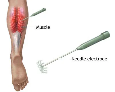
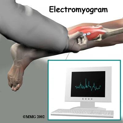
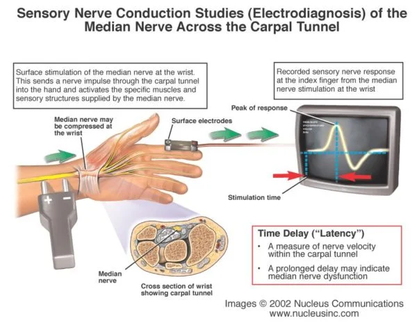
Electrodiagnostics (EDX) testing is used to evaluate the integrity and function of the peripheral nervous system (most cranial nerves, spinal roots, plexi, and nerves), NMJ, muscles, and the central nervous system (brain and spinal cord). EDX testing is performed as part of an EDX consultation for diagnosis or as follow-up of an existing condition. EDX studies can provide information to: 

  • Identify normal and abnormal nerve, muscle, motor or sensory neuron, and NMJ functioning.
  • Localize region(s) of abnormal function.
  • Define the type of abnormal function.
  • Determine the distribution of abnormalities.
  • Determine the severity of abnormalities.
  • Estimate the date of a specific nerve injury.
  • Estimate the duration of the disease.
  • Determine the progression of abnormalities or of recovery from abnormal function.
  • Aid in diagnosis and prognosis of disease.
  • Aid in selecting treatment options.
  • Assists in following response to treatment by providing objective evidence of change in neuromuscular function.
  • Localize correct locations for injection of intramuscular agents (e.g., botulinum toxin).
Discogram Testing by Padda Institute
Electromyography (EMG) is a technique for evaluating and recording the electrical activity produced by skeletal muscles. An EMG detects the electrical potential generated by muscle cells when electrically or neurologically activated. Often, EMG testing is performed with another test that measures the conducting function of nerves, called a nerve conduction study (NCS). These electrodiagnostics tests (EMG and NCS) are often performed at the same office visit and by the same personnel, the risks and procedures generally apply to both tests.
EMG Testing and Results - Padda Institute
In some medical conditions the electrical activity of the muscles or nerves is not normal. Finding and describing these electrical properties in the muscle or nerve may help diagnose your condition. EDX may aid with the diagnosis of nerve compression or injury (such as carpal tunnel syndrome), nerve root injury (such as sciatica), and with other problems of the muscles or nerves. Less common medical conditions include amyotrophic lateral sclerosis, myasthenia gravis, and muscular dystrophy.
EMG is most often used when people have symptoms of weakness, and examination shows impaired muscle strength. It can help to tell the difference between muscle weakness caused by injury of a nerve attached to a muscle and weakness due to neurologic disorders.
Dorsal (back) of spinal cord transmits sensory information from sensory units to brain, Ventral (front) of cord transmits motor information from brain to muscle

Risks

People usually have a small amount of discomfort during EMG testing because of pin insertion. Disposable needles are used so there is no risk of infection.
During nerve conduction studies, small electrodes are taped to the skin or placed around fingers. You typically experience a brief and mild shock, which may be a bit unpleasant. Most people find it only slightly annoying.
It is very important to note that most EMG/NCV tests are not 100% accurate. Most physicians will admit that the tests have at least a 10% margin of error. Very often individuals with nerve damage will have normal EMG/NCV tests even though they are experiencing nerve damage.
Electrode Needles - Electrodiagnostics Testing

During The Procedure

During EMG, small pins or needles are inserted into muscles to measure electrical activity. The needles are different than needles used for injection of medications. They are small and solid, not hollow like hypodermic needles. Because no medication is injected, discomfort is much less than with shots.
You will be asked to contract your muscles by moving a small amount during the testing.
With nerve conduction studies, small electrodes will be taped to your skin or placed around your fingers. You typically will experience a mild and brief tingling or shock, which may be a bit unpleasant.
The person who administers the test will explain the procedure. Often muscle activity is monitored through a speaker during the test, which may make a popping or soft roaring noise. The EMG technician will be looking at an oscilloscope, which looks like a small TV set during the procedure.
Electrical impulses are recorded for detailed analysis after the EMG test is completed.
Electromyography Procedure pa

How Long Does EDX Take?

Testing may take 30-60 minutes. The nerve conduction part of the test usually takes longer than the needle exam because one needs to make calculations and measurements during it. On average, if one extremity is studied, the nerve conductions take anywhere between 15 and 30 minutes. The needle exam for one extremity usually takes 15 to 20 minutes. You can count on being in the examination room for about one hour if only one extremity is requested; longer if more extremities need to be tested.

Carpal Tunnel Syndrome

For suspected carpal tunnel syndrome (CTS), bilateral median motor and sensory NCSs are often indicated. The studies in the contralateral asymptomatic limb serve as controls in cases where values are borderline and may establish the presence of bilateral CTS, which is a frequent finding.
Two to 4 additional sensory or mixed NCSs can be compared to the median sensory NCSs to increase the diagnostic sensitivity of the testing. The additional sensory NCSs and an additional motor NCS (usually ulnar) are indicated to exclude a generalized neuropathy or multiple mononeuropathies.
If 2 sensitive sensory NCSs are performed to start, additional sensory testing on the same limb is rarely needed. For suspected bilateral CTS, bilateral median motor and sensory NCSs are indicated. Up to 2 additional motor and 2 additional sensory NCSs are often indicated. The extent of the needle EMG examination depends on the results of the NCSs and the differential diagnosis considered in the individual patient.
About Carpal Tunnel Syndrome

Radiculopathy

A minimal evaluation for radiculopathy includes 1 motor and 1 sensory NCS and a needle EMG examination of the involved limb. However, the EDX testing can include up to 3 motor NCSs (in cases of an abnormal motor NCS, the same nerve in the contralateral limb and another motor nerve in the ipsilateral limb can be studied) and 2 sensory NCSs. Bilateral studies are often necessary to exclude a central disc herniation with bilateral radiculopathies or spinal stenosis or to differentiate between radiculopathy and plexopathy, polyneuropathy, or mononeuropathy.
H reflexes and F waves can provide useful complementary information that is helpful in the evaluation of suspected radiculopathy and can add to the certainty of electrodiagnostic information supporting a diagnosis of root dysfunction.
Additional testing may be indicated in patients with a differential diagnosis which includes peripheral neuropathy, cervical radiculopathy, brachial plexopathy, or more proximal median neuropathy.

Mononeuropathy and Polyneuropathy or Mononeuropathy Multiplex

Mononeuropathy and polyneuropathy/mononeuropathy multiplex are entirely different conditions and must be considered separately. Mononeuropathy comprises focal lesions of a single peripheral nerve. Polyneuropathy comprises diseases in which there is a bilaterally symmetric disturbance of peripheral nerve functions. Mononeuropathy multiplex comprises multifocal isolated lesions of more than 1 peripheral nerve.

Mononeuropathy

To determine the level of the lesion in a focal mononeuropathy, and in order to exclude radiculopathy, plexopathy, or polyneuropathy, it may be necessary to study 3 motor and 3 sensory nerves including the clinically affected nerve, the same nerve on the contralateral side, and an unaffected ipsilateral nerve. F-wave studies provide additional diagnostic information. A needle EMG examination in the affected limb is indicated.

Polyneuropathy or Mononeuropathy Multiplex

In order to characterize the nature of the polyneuropathy (axonal or demyelinating, diffuse or multifocal) and in order to exclude polyradiculopathy, plexopathy, neuronopathy, or multiple mononeuropathies, it may be necessary to study 4 motor and 4 sensory nerves, consisting of 2 motor and 2 sensory NCSs in 1 leg, 1 motor and 1 sensory NCS in the opposite leg, and 1 motor and 1 sensory NCS in 1 arm. H-reflex studies and F-wave studies from 2 nerves may provide additional diagnostic information. At least 2 limbs should be studied by a needle EMG examination. Studies of related paraspinal muscles are indicated to exclude some conditions such as polyradiculopathy.

Myopathy

To diagnose a myopathy, a needle EMG examination of 2 limbs is indicated. To help exclude other disorders such as polyneuropathy or neuronopathy, 2 motor and 2 sensory NCSs are indicated. Two repetitive motor nerve stimulation studies may be performed to exclude a disorder of neuromuscular transmission.

Motor Neuronopathy

In order to establish the diagnosis of motor neuronopathy (for example, amyotrophic lateral sclerosis [ALS or Lou Gehrig’s disease]) and to exclude other disorders in the differential diagnosis, such as multifocal motor neuropathy or polyneuropathy, up to 4 motor nerves and 2 sensory nerves may be studied. Needle EMG of up to 4 extremities (or 3 limbs and facial or tongue muscles) is often necessary to document widespread denervation and to exclude a myopathy. One repetitive motor nerve stimulation study may be indicated to exclude a disorder affecting neuromuscular transmission.

Plexopathy

To characterize a brachial plexopathy and to differentiate it from cervical radiculopathy and mononeuropathies, it is often necessary to study all major sensory and motor nerves that can be easily studied in both upper extremities (radial, median, ulnar, and medial and lateral antebrachial cutaneous sensory; radial, median, ulnar, and possibly axillary and musculocutaneous motor) and to perform a needle EMG examination in both upper extremities. To characterize the lumbosacral plexopathy and to differentiate it from lumbar radiculopathy and mononeuropathies, it is often necessary to study all major sensory and motor nerves that can be easily studied in both lower extremities (superficial peroneal and sural sensory; peroneal and posterior tibial motor) and to perform a needle EMG examination in both lower extremities. F-wave studies in the motor nerves and soleus H reflexes also provide useful information.

Neuromuscular Junction

To demonstrate and characterize abnormal neuromuscular transmission, repetitive nerve stimulation studies should be performed in up to 2 nerves and SFEMG in up to 2 muscles. If any of these are abnormal, up to 2 motor and 2 sensory NCSs may be performed to exclude neuropathies that can be associated with abnormal neuromuscular transmission. At least 1 motor and 1 sensory NCS should be performed in a clinically involved limb, preferably in the distribution of a nerve studied with repetitive stimulation or SFEMG. At least 1 distal and 1 proximal muscle should be studied by a needle EMG examination to exclude a neuropathy or myopathy that can be associated with abnormal repetitive stimulation studies or SFEMG. At least 1 of the muscles should be clinically involved and both muscles should be in clinically involved limbs.

Timing of Testing After an Injury

In combination, NCSs and a needle EMG examination may be most helpful when performed several weeks after the injury has occurred. However, NCSs are often useful acutely after nerve injury, for example, if there is concern that a nerve has been severed. In fact, if studies are delayed, the opportunity to precisely identify the region of injury or to intervene may be lost. In some cases, even needle EMG testing performed immediately after a nerve injury may demonstrate abnormal motor unit action potential (MUAP) recruitment and/or provide baseline information that can be helpful in documenting preexisting conditions, date the injury, or serve as a baseline for comparison with later studies.
Because of the variability of different nerve injuries, a standard rule on the timing of EDX testing cannot easily be established and the AAEM does not have specific recommendations in this regard. In all instances, the AAEM encourages dialogue between physicians and payors and encourages the appropriate use of the physician’s clinical judgment in determining when studies are most appropriately performed and what studies should be conducted.

Get Answers with Electrodiagnostics Nerve Tests

Contact us today to schedule an Electrodiagnostic study and take the first step toward understanding and addressing your nerve concerns.

Frequency Of Electrodiagnostic Testing In A Given Patient

There are many clinical situations where good medical management requires repeat testing, such as in the following examples: 

  • Second diagnosis. Where a single diagnosis is made on the first visit, but the patient subsequently develops a new set of symptoms, further evaluation is required for a second diagnosis.
  • Inconclusive diagnosis. When a serious diagnosis (e.g., ALS) is suspected but the results of the needle EMG/NCS examination are insufficient to be conclusive, 1, or even more, follow-up studies are needed to establish or exclude the diagnosis.
  • Rapidly evolving disease. Initial EDX testing in some diseases may not show any abnormality (e.g., Guillain-Barré syndrome) in the first 1 to 2 weeks. An early diagnosis confirmed by repeat electrodiagnosis must be made quickly so that treatment can begin. Follow-up testing can be extremely useful in establishing prognosis and monitoring patient status.
  • Course of the disease. Certain treatable diseases such as polymyositis and myasthenia gravis follow a fluctuating course with variable response to treatment. The physician treating such patients needs to monitor the disease progress and the response to therapeutic interventions. The results of follow-up evaluations may be necessary to guide treatment decisions.
  • Unexpected course or change in course of the disease. In certain situations, management of a diagnosed condition may not yield expected results or new, questionably related problems may occur (e.g., failure to improve following surgery for radiculopathy). In these instances, reexamination is appropriate.
  • Recovery from injury. Repeat evaluations may be needed to monitor recovery, to help establish prognosis, and/or to determine the need for and timing of surgical intervention (e.g., traumatic nerve injury).

Repeat EDX consultation is therefore sometimes necessary and, when justifiable, should be reimbursed. Reasonable limits can be set concerning the frequency of repeat EDX testing per year in a given patient by a given EDX consultant for a given diagnosis. The following numbers of tests per 12-month period per diagnosis per physician are acceptable:

  • Two tests for carpal tunnel-unilateral, carpal tunnel-bilateral, radiculopathy, mononeuropathy, polyneuropathy, myopathy, and NMJ disorders.
  • Three tests for motor neuronopathy and plexopathy.

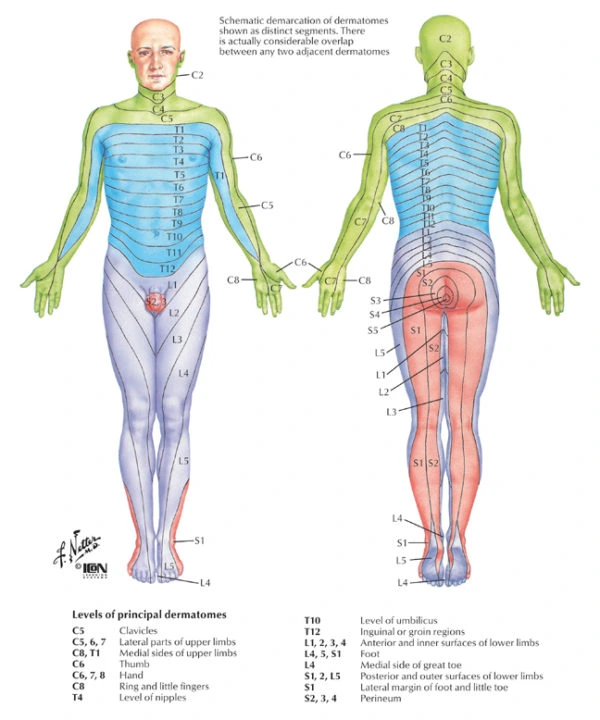
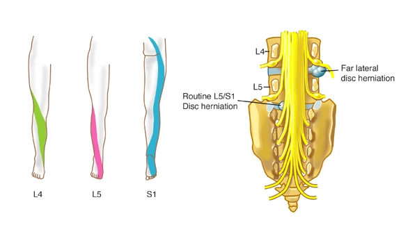
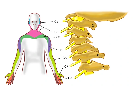
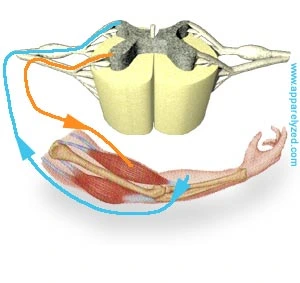
Spinal nerves have motor fibers and sensory fibers. The motor fibers innervate certain muscles, while the sensory fibers innervate certain areas of skin. A skin area innervated by the sensory fibers of a single nerve root is known as a dermatome. A group of muscles primarily innervated by the motor fibers of a single nerve root is known as a myotome. Although slight variations do exist, dermatome and myotome patterns of distribution are relatively consistent from person to person.

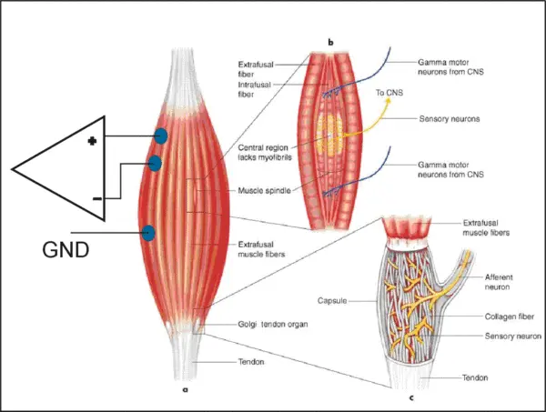
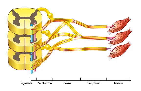
The ventral (anterior) gray matter of the spinal cord contains nerve cells that send axon fibers out, through the nerves, to their end points on the muscles that they activate. Sensory information from the body and arriving instructions from the brain all cause movement by giving instructions to these “motor neurons” in the spinal cord gray matter.

Spinal Cord Segmental Myotomes And Dermatomes

Myotomes – Relationship between the spinal nerve & muscle and are best evaluated with EMG Dermatomes – Relationship between the spinal nerve & skin and a combination of EMG and NCS is used to define pathology.

Myotomes

Each muscle in the body is supplied by a particular level or segment of the spinal cord and by its corresponding spinal nerve. The muscle, and its nerve make up a myotome. This is approximately the same for every person and are as follows: 

  • C3,4 and 5 supply the diaphragm (the large muscle between the chest and the belly that we use to breath).
  • C5 also supplies the shoulder muscles and the muscle that we use to bend our elbow
  • C6 is for bending the wrist back.
  • C7 is for straightening the elbow.
  • C8 bends the fingers.
  • T1 spreads the fingers.
  • T1 –T12 supplies the chest wall & abdominal muscles.
  • L2 bends the hip.
  • L3 straightens the knee.
  • L4 pulls the foot up.
  • L5 wiggles the toes.
  • S1 pulls the foot down.
  • S3,4 and 5 supply the bladder. bowel and sex organs and the anal and other pelvic muscles.
Sensory dermatomes in blue, motor myotomes in yeallow
Myotome Muscle EMG
Myotome muscle EMG
Spinal Cord Segmental Dermatomes

Spinal Cord Segmental Dermatomes

Dermatome is a Greek word which literally means “skin cutting”. A dermatome is an area of the skin supplied by nerve fibers originating from a single dorsal nerve root. The dermatomes are named according to the spinal nerve which supplies them. The dermatomes form into bands around the trunk but in the limbs their organisation is more complex as a result of the dermatomes being “pulled out” as the limb buds form and develop into the limbs during embryological development.
In diagrams or maps, the boundaries of dermatomes are usually sharply defined. However, in life there is considerable overlap of innervation between adjacent dermatomes. Thus, if there is a loss of afferent nerve function by one spinal nerve sensation from the region of skin which it supplies is not usually completely lost as overlap from adjacent spinal nerves occurs: however, there will be a reduction in sensitivity.
Different parts of the spinal cord provide innervation to the body, organized as dermatomes.
Lumbar Radiculopathy - Pain Treatment Center in St. Louis
Cervical Radiculopathy - Electrodiagnostics Testing

Send A Message

For questions and concerns, please fill out the form below.

Pain Conditions We Treat

At Padda Institute, our pain management specialists & pain specialist doctors provide you the guidance and treatments tailored to your needs. We are best at what we do; we diagnose and evaluate to determine your best treatment plan, so that you can experience chronic pain relief like never before.