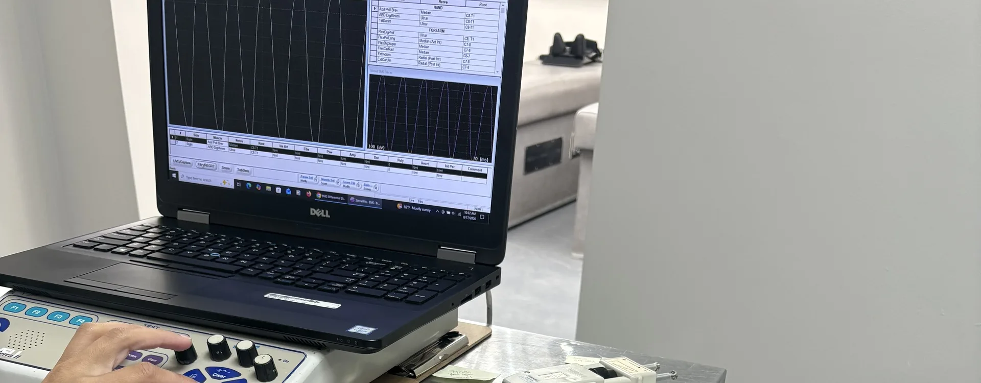



EMG and nerve conduction study console showing live nerve traces and the muscle, nerve and root worksheet on screen at Padda Institute, St. Louis

EMG nerve testing · St. Louis

Electrodiagnostics

Electrodiagnostic testing is an EMG plus a nerve conduction study. It records the electrical activity of your muscles and nerves. It finds where a nerve is damaged and how badly. It diagnoses a pinched nerve such as carpal tunnel syndrome, nerve root injury such as sciatica, and other muscle and nerve problems.

Take the first step toward relief. Fill out the form, and we’ll reach out for a private talk about your needs.

Prefer not to fill in a form?

Same-day and emergency appointments are available.

What is electrodiagnostic (EMG and nerve conduction) testing?

Electrodiagnostics (EDX) testing checks the health and function of the peripheral nervous system. That covers most cranial nerves, spinal roots, plexi (nerve networks), and nerves. It also checks the NMJ (the joint where nerve meets muscle), the muscles, and the central nervous system (brain and spinal cord). We do EDX testing as part of an EDX visit, to make a diagnosis or to follow a known condition. EDX studies can help:

  • Tell normal from abnormal function in nerves, muscles, motor or sensory nerve cells, and the NMJ.
  • Find the area(s) that are not working right.
  • Name the type of problem.
  • Show how far the problem spreads.
  • Show how bad the problem is.
  • Estimate when a nerve injury happened.
  • Estimate how long the disease has been there.
  • Show whether the problem is getting worse or healing.
  • Help diagnose disease and predict its course.
  • Help pick treatment.
  • Track your response to treatment with hard proof of change in nerve and muscle function.
  • Find the right spots to inject medicine into muscle (e.g., botulinum toxin).

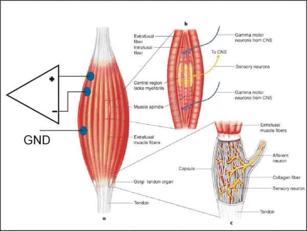
Electromyography (EMG) is a test that checks and records the electrical activity made by your muscles. An EMG picks up the electrical charge muscle cells make when a nerve or a small current fires them. EMG is often done with a second test that measures how well nerves carry signals. That test is called a nerve conduction study (NCS). These two tests (EMG and NCS) are often done at the same visit by the same staff. The risks and steps mostly apply to both.

Numbness and weakness are only part of what raises the risk of a fall. If you are also unsteady, we read electrodiagnostic testing next to balance and fall-risk testing. That testing measures the inner ear, thinking, and walking, as well as the nerves.

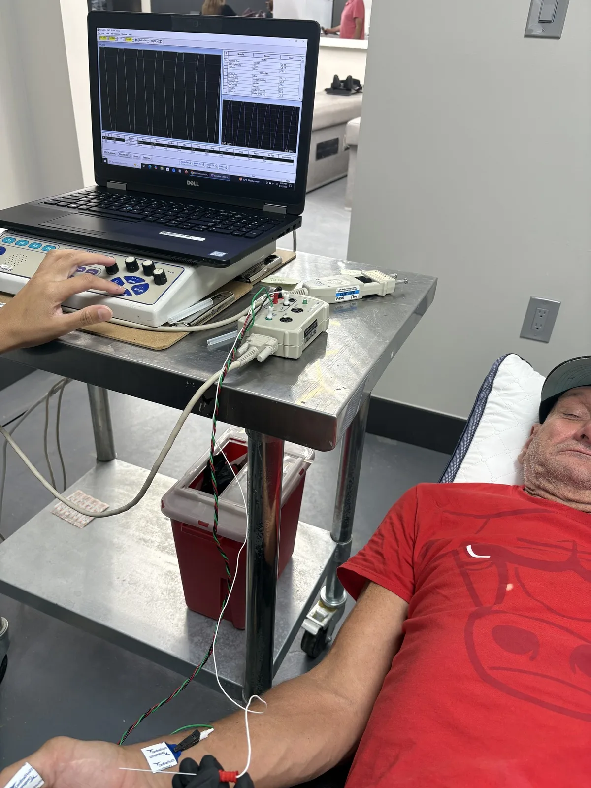
Technician operating the EMG recording console while a patient rests with nerve conduction electrodes taped to the forearm and hand at Padda Institute, St. Louis
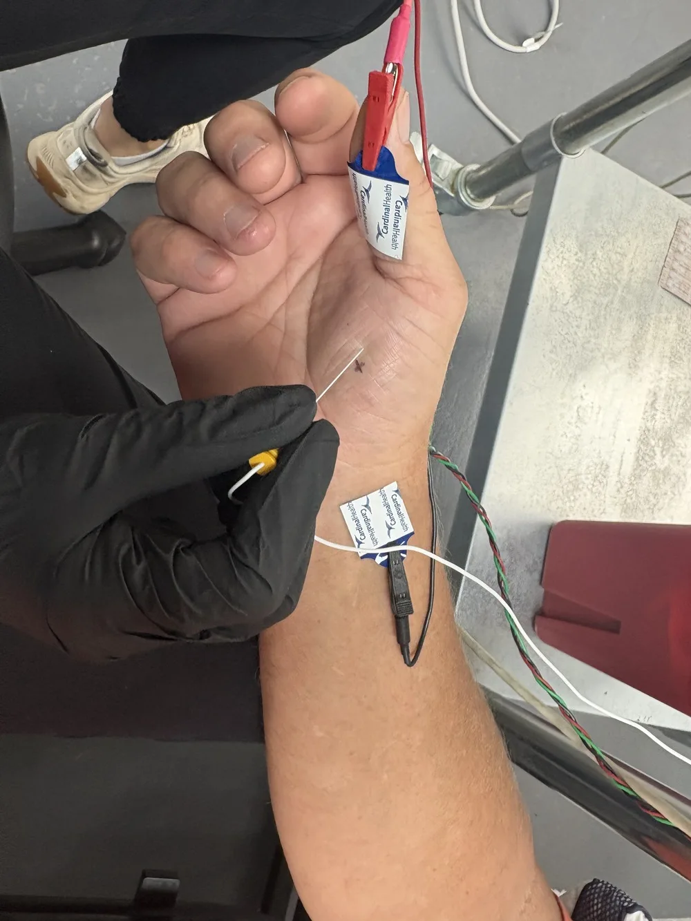
Close view of recording electrodes taped over the thenar eminence with a wrist reference electrode in place before median nerve stimulation at Padda Institute, St. Louis

In some conditions, the electrical activity of the muscles or nerves is not normal. Finding and describing that activity can help diagnose your condition. EDX can help diagnose a pinched or injured nerve (such as carpal tunnel syndrome). It can help with nerve root injury (such as sciatica) and other muscle or nerve problems. Less common conditions include amyotrophic lateral sclerosis, myasthenia gravis, and muscular dystrophy.

EMG is most often used when people feel weak and the exam shows reduced muscle strength. It can help tell apart two kinds of weakness. One comes from an injured nerve that runs to a muscle. The other comes from a neurologic disorder (a disease of the brain or nervous system).

Dorsal (back) of spinal cord transmits sensory information from sensory units to brain, Ventral (front) of cord transmits motor information from brain to muscle

Does an EMG test hurt, and how accurate is it?

Most people feel a little discomfort during EMG testing when the pins go in. We use throwaway needles, so there is no risk of infection.

During nerve conduction studies, small electrodes are taped to the skin or placed around fingers. You will usually feel a quick, mild shock. It may be a bit unpleasant. Most people find it only a little annoying.

Know this: most EMG/NCV tests are not 100% accurate. Most physicians will admit the tests have at least a 10% margin of error. Very often, people with real nerve damage still get normal EMG/NCV results.

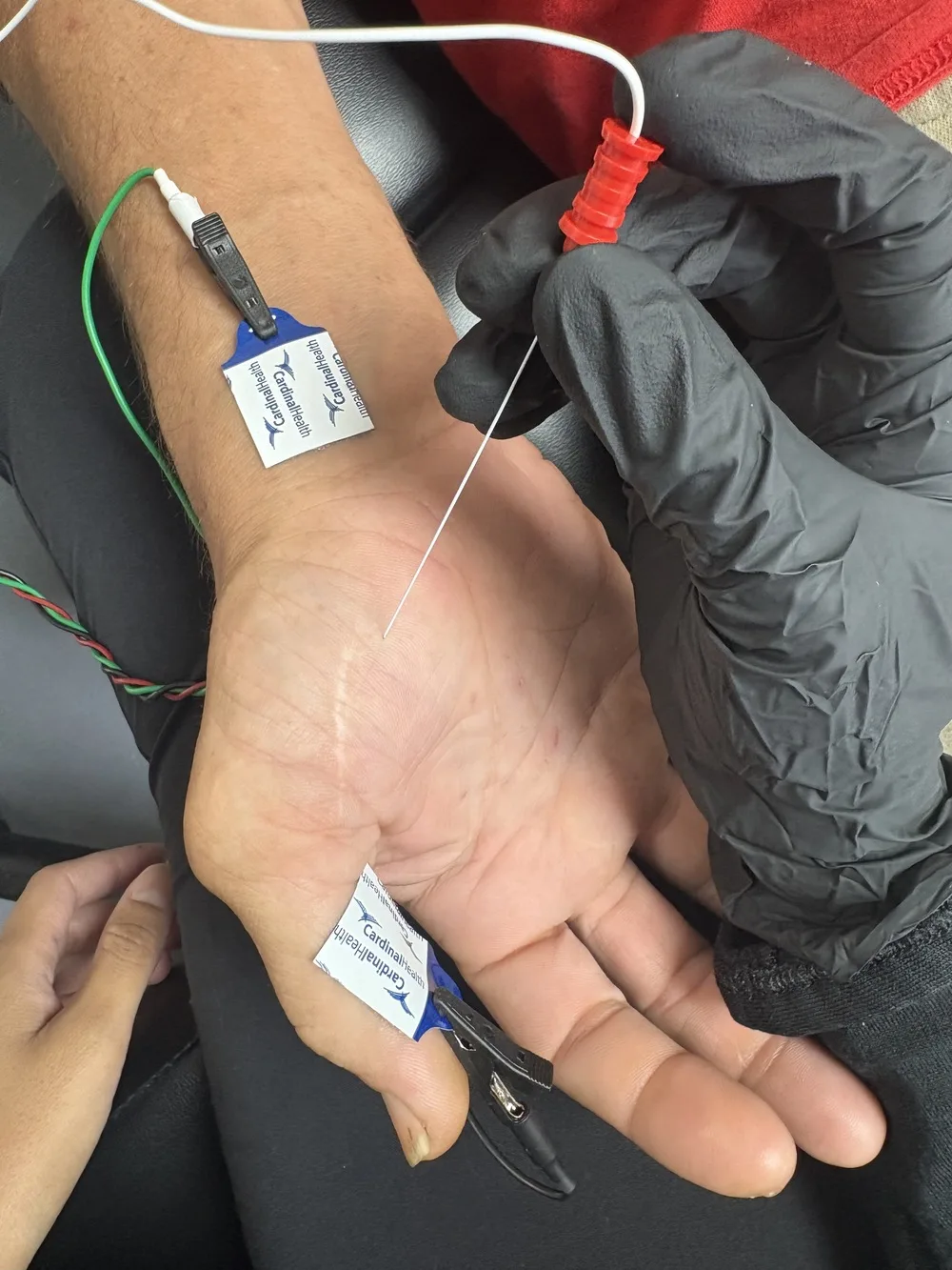
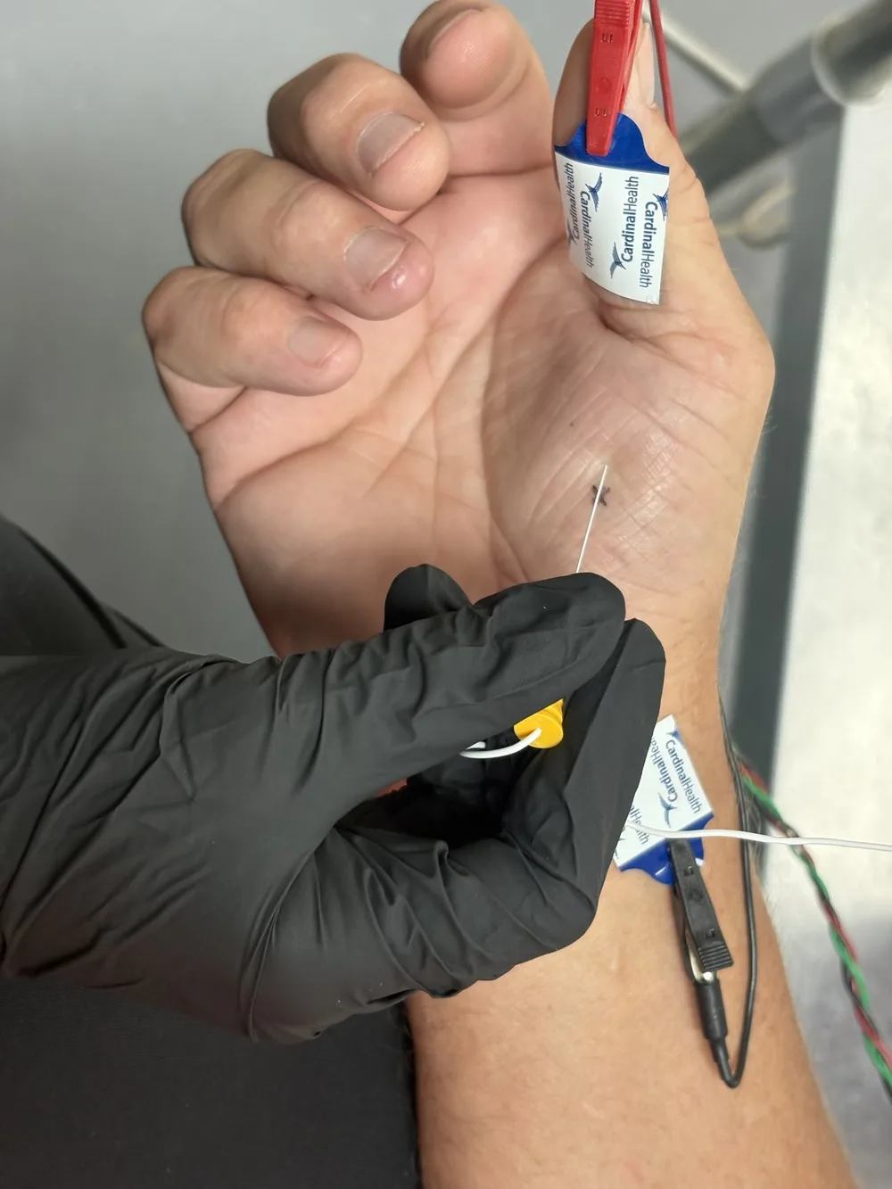
Gloved clinician holding a stimulating probe against the palm with surface recording electrodes taped to the hand and wrist during a nerve conduction study at Padda Institute, St. Louis

What happens during an EMG and nerve conduction study?

During EMG, small pins or needles go into muscles to measure electrical activity. These are not the needles used to inject medicine. They are small and solid, not hollow like a shot needle. No medicine goes in, so it hurts much less than a shot.

You will be asked to tighten your muscles by moving a little during the test.

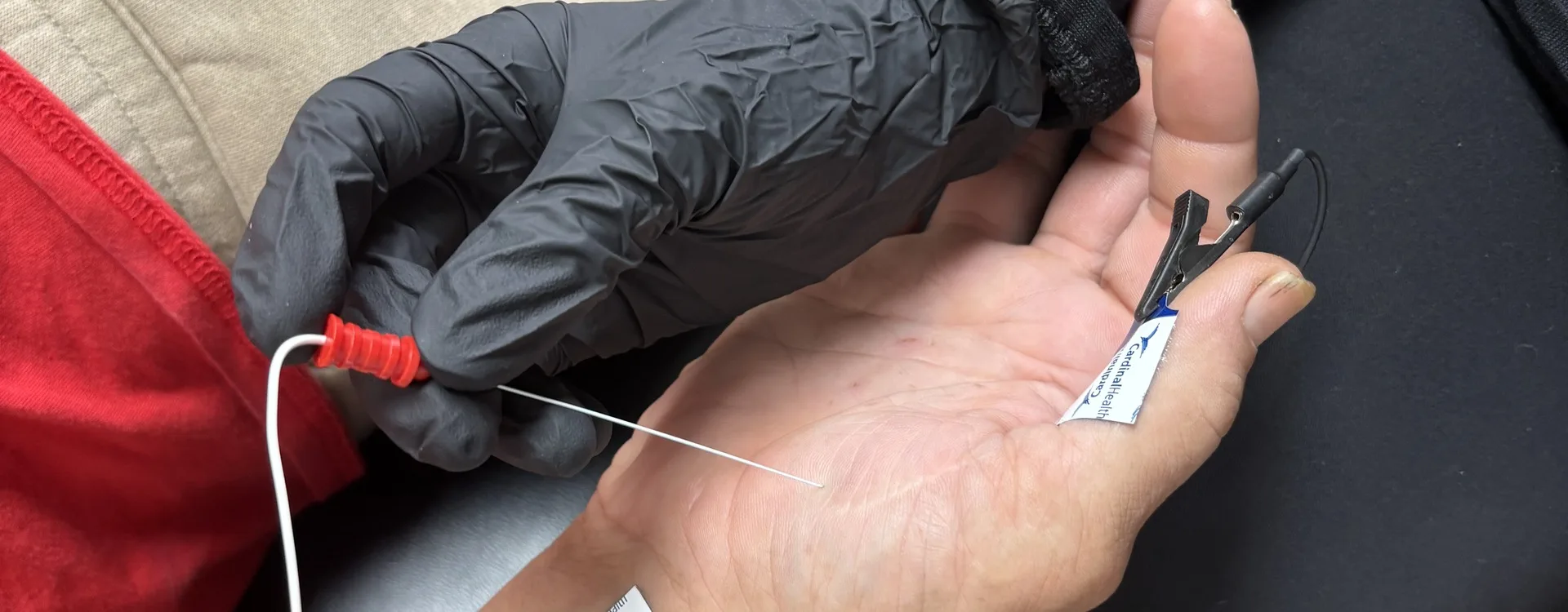
With nerve conduction studies, small electrodes will be taped to your skin or placed around your fingers. You will usually feel a mild, quick tingle or shock. It may be a bit unpleasant.

The person giving the test will explain each step. Often you can hear muscle activity through a speaker during the test. It may make a popping or soft roaring sound. The EMG technician will watch an oscilloscope, a screen that looks like a small TV.

The electrical signals are recorded so we can study them in detail after the test.

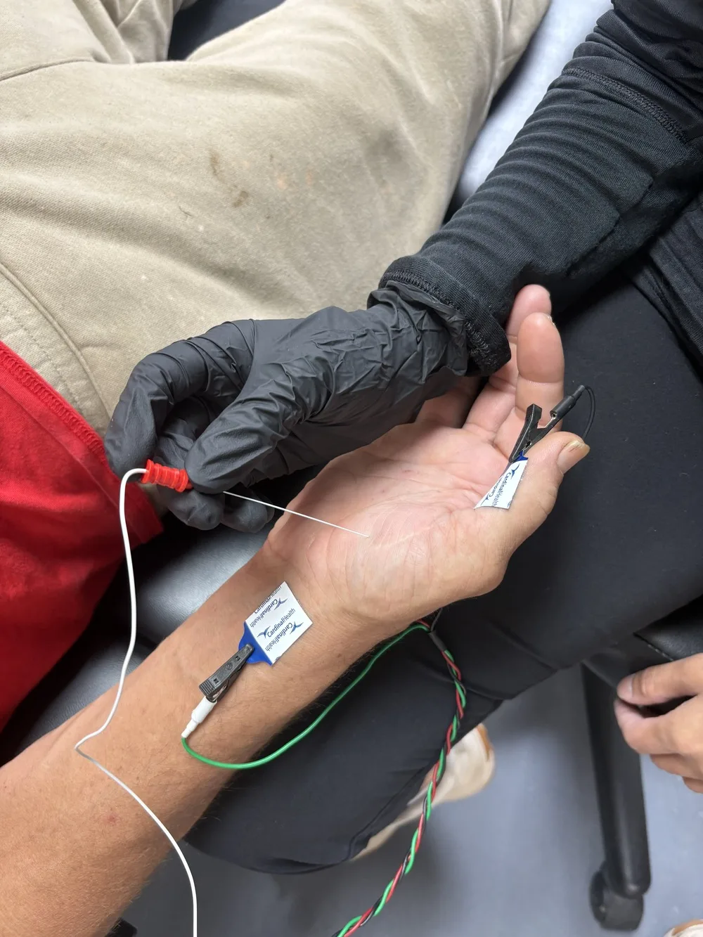
Stimulating probe held at the wrist crease with recording electrodes on the thumb and forearm during median nerve conduction testing at Padda Institute, St. Louis

How long does an EMG and nerve conduction test take?

Testing may take 30-60 minutes. The nerve conduction part usually takes longer than the needle exam. That is because we take measurements and do math during it. On average, for one arm or leg, the nerve conduction part takes 15 to 30 minutes. The needle exam for one arm or leg usually takes 15 to 20 minutes. Plan on about one hour in the exam room if only one limb is tested. It takes longer if more limbs need testing.

Carpal Tunnel Syndrome

For suspected carpal tunnel syndrome (CTS), we often test the median nerve in both hands, for both strength and feeling (bilateral median motor and sensory NCSs). The hand without symptoms serves as a control when values are borderline. It may also show CTS in both hands, which is common.

Two to 4 more sensory or mixed NCSs can be compared to the median sensory NCSs. That makes the test better at catching the problem. The extra sensory NCSs and one more motor NCS (usually ulnar) help rule out nerve damage all over the body, or damage to several single nerves.

If 2 sensitive sensory NCSs are done first, more sensory testing on the same limb is rarely needed. For suspected CTS in both hands, bilateral median motor and sensory NCSs are called for. Up to 2 more motor and 2 more sensory NCSs are often called for. How much needle EMG we do depends on the NCS results and on what else could be causing the problem in that patient.

Median nerve stimulation at the wrist with recording electrodes on the thumb and forearm, the study used to separate carpal tunnel syndrome from cervical radiculopathy at Padda Institute, St. Louis

Radiculopathy

The least testing for radiculopathy (a pinched nerve root) is 1 motor and 1 sensory NCS plus a needle EMG of the problem limb. But EDX testing can include up to 3 motor NCSs and 2 sensory NCSs. If a motor NCS is abnormal, we can test the same nerve on the other side and another motor nerve on the same side. Testing both sides is often needed. It rules out a central disc herniation pinching roots on both sides, or spinal stenosis. It also tells radiculopathy apart from plexopathy, polyneuropathy, or mononeuropathy.

H reflexes and F waves can add useful information when we suspect radiculopathy. They can make us more certain the nerve root is the problem.

More testing may be needed when the list of possible causes includes peripheral neuropathy, cervical radiculopathy, brachial plexopathy, or median nerve damage higher up the arm.

Mononeuropathy and Polyneuropathy or Mononeuropathy Multiplex

When one nerve is damaged (focal mononeuropathy), we need to find where along it the damage sits. We also need to rule out radiculopathy, plexopathy, or polyneuropathy. That may take testing 3 motor and 3 sensory nerves. They include the problem nerve, the same nerve on the other side, and a healthy nerve on the same side. F-wave studies add more information. A needle EMG of the problem limb is called for.

Polyneuropathy or Mononeuropathy Multiplex

Polyneuropathy is damage to many nerves. To pin down its type, we ask two things. Is the nerve core (axonal) or the nerve coating (demyelinating) damaged? Is it spread out (diffuse) or in patches (multifocal)? We also need to rule out polyradiculopathy, plexopathy, neuronopathy, or multiple mononeuropathies. That may take testing 4 motor and 4 sensory nerves. That means 2 motor and 2 sensory NCSs in 1 leg, 1 motor and 1 sensory NCS in the other leg, and 1 motor and 1 sensory NCS in 1 arm. H-reflex and F-wave studies from 2 nerves may add more information. At least 2 limbs should get a needle EMG. Testing the muscles next to the spine helps rule out some conditions such as polyradiculopathy.

Myopathy

To diagnose a myopathy (a muscle disease), we do a needle EMG of 2 limbs. To help rule out other problems such as polyneuropathy or neuronopathy, we do 2 motor and 2 sensory NCSs. Two repeated motor nerve stimulation studies may be done. They rule out a problem where nerve signals fail to cross to the muscle.

Motor Neuronopathy

Motor neuronopathy is disease of the nerve cells that run the muscles (for example, amyotrophic lateral sclerosis [ALS or Lou Gehrig’s disease]). To diagnose it, we must rule out look-alikes such as multifocal motor neuropathy or polyneuropathy. We may test up to 4 motor nerves and 2 sensory nerves. Needle EMG of up to 4 limbs (or 3 limbs and face or tongue muscles) is often needed. It shows whether muscles have lost their nerve supply in many places. It also rules out a myopathy. One repeated motor nerve stimulation study may be needed to rule out a problem where nerve signals fail to cross to the muscle.

Plexopathy

A brachial plexopathy is damage to the nerve network in the shoulder. To describe it and tell it apart from cervical radiculopathy and mononeuropathies, we often test every major nerve that is easy to test in both arms. That means the sensory nerves (radial, median, ulnar, and medial and lateral antebrachial cutaneous sensory). It also means the motor nerves (radial, median, ulnar, and possibly axillary and musculocutaneous motor). We also do a needle EMG in both arms. A lumbosacral plexopathy is damage to the nerve network in the low back and pelvis. To describe it and tell it apart from lumbar radiculopathy and mononeuropathies, we often test every major nerve that is easy to test in both legs (superficial peroneal and sural sensory; peroneal and posterior tibial motor). We also do a needle EMG in both legs. F-wave studies in the motor nerves and soleus H reflexes add useful information too.

Neuromuscular Junction

Sometimes nerve signals fail to cross from nerve to muscle. To show and describe that, we do repeated nerve stimulation studies in up to 2 nerves and SFEMG (single-fiber EMG) in up to 2 muscles. If any of these are abnormal, we may do up to 2 motor and 2 sensory NCSs. Those rule out nerve diseases that can come with this signal problem. At least 1 motor and 1 sensory NCS should be done in a limb with symptoms. Ideally it is in the area of a nerve tested with repeated stimulation or SFEMG. At least 1 muscle far from the trunk and 1 muscle close to it should get a needle EMG. That rules out a nerve or muscle disease that can come with abnormal repeated stimulation studies or SFEMG. At least 1 of the muscles should have symptoms, and both muscles should be in limbs with symptoms.

Timing of Testing After an Injury

Together, NCSs and a needle EMG may help most when done several weeks after the injury. But NCSs are often useful right after a nerve injury. One example is when a nerve may have been cut. In fact, if testing waits, the chance to find the exact spot of injury, or to act on it, may be lost. In some cases, even a needle EMG done right after a nerve injury can show abnormal motor unit action potential (MUAP) recruitment. It can also give a starting point. That helps record problems you already had, date the injury, or compare with later tests.

Nerve injuries vary a lot. So there is no simple rule on when to do EDX testing, and the AAEM has no specific advice on it. In every case, the AAEM urges physicians and payors to talk. It also urges physicians to use their own clinical judgment on when to test and which tests to do.

Gloved clinician holding the stimulating probe against a patient's palm with a recording electrode clipped to the finger during a nerve conduction study at Padda Institute, St. Louis

Get Answers with Electrodiagnostics Nerve Tests

Contact us today to schedule an Electrodiagnostic study. Take the first step toward understanding and addressing your nerve problem.

When is a repeat EMG test needed?

Good medical care often calls for repeat testing. Here are some examples:

  • Second diagnosis. One diagnosis is made on the first visit. Later, the patient gets a new set of symptoms. More testing is needed for a second diagnosis.
  • Unclear diagnosis. A serious disease (e.g., ALS) is suspected, but the needle EMG/NCS results are not enough to be sure. Then 1, or even more, follow-up studies are needed to prove or rule out the diagnosis.
  • Fast-moving disease. In some diseases, the first EDX test may look normal (e.g., Guillain-Barré syndrome) in the first 1 to 2 weeks. An early diagnosis, proven by a repeat test, must come fast so treatment can start. Follow-up testing can be very useful to predict the course and watch how the patient is doing.
  • Course of the disease. Some treatable diseases, such as polymyositis and myasthenia gravis, go up and down. Their response to treatment varies. The physician treating these patients needs to track the disease and the response to treatment. Follow-up test results may be needed to guide treatment choices.
  • Unexpected course or change in course of the disease. Sometimes treating a known condition does not give the results we expect. Or new problems show up that may or may not be related (e.g., no improvement after surgery for radiculopathy). In these cases, testing again makes sense.
  • Recovery from injury. Repeat tests may be needed to track healing, to help predict the outcome, and/or to decide whether and when surgery is needed (e.g., traumatic nerve injury).

So a repeat EDX visit is sometimes needed. When it is justified, insurance should pay for it. Fair limits can be set on how often one EDX doctor repeats testing in one patient for one diagnosis each year. These numbers of tests per 12-month period per diagnosis per physician are acceptable:

  • Two tests for carpal tunnel-unilateral, carpal tunnel-bilateral, radiculopathy, mononeuropathy, polyneuropathy, myopathy, and NMJ disorders.
  • Three tests for motor neuronopathy and plexopathy.

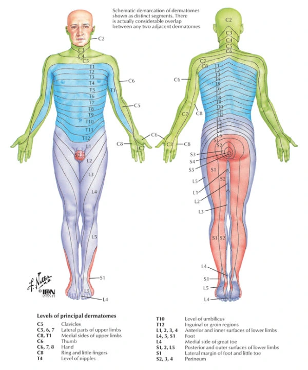
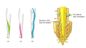
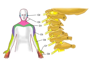
Spinal nerves have motor fibers and sensory fibers. The motor fibers run certain muscles. The sensory fibers serve certain areas of skin. A patch of skin served by the sensory fibers of one nerve root is called a dermatome. A group of muscles run mainly by the motor fibers of one nerve root is called a myotome. There are small differences, but dermatome and myotome maps are fairly steady from person to person.

The front (ventral, or anterior) gray matter of the spinal cord holds nerve cells. They send long fibers out, through the nerves, to the muscles they fire. Feeling from the body and orders from the brain both cause movement. They do it by giving orders to these “motor neurons” in the spinal cord gray matter.

Spinal Cord Segmental Myotomes And Dermatomes

Myotomes – the link between the spinal nerve & muscle. EMG checks them best. Dermatomes – the link between the spinal nerve & skin. A mix of EMG and NCS is used to find the problem.

Myotomes

Each muscle in the body is run by one level of the spinal cord and its matching spinal nerve. The muscle and its nerve make up a myotome. This is about the same for every person:

  • C3,4 and 5 supply the diaphragm (the large muscle between the chest and the belly that we use to breathe).
  • C5 also supplies the shoulder muscles and the muscle that we use to bend our elbow.
  • C6 is for bending the wrist back.
  • C7 is for straightening the elbow.
  • C8 bends the fingers.
  • T1 spreads the fingers.
  • T1 –T12 supplies the chest wall & abdominal muscles.
  • L2 bends the hip.
  • L3 straightens the knee.
  • L4 pulls the foot up.
  • L5 wiggles the toes.
  • S1 pulls the foot down.
  • S3,4 and 5 supply the bladder, bowel and sex organs and the anal and other pelvic muscles.
Sensory dermatomes in blue, motor myotomes in yellow
EMG Muscle Configuration
Myotome muscle EMG
Electrodiagnostics

Spinal Cord Segmental Dermatomes

Dermatome is a Greek word which literally means “skin cutting”. A dermatome is an area of skin served by nerve fibers from one dorsal nerve root. Each dermatome is named for the spinal nerve that serves it. Around the trunk, dermatomes form bands. In the arms and legs, the layout is more complex. That is because the dermatomes get “pulled out” as the limb buds grow into limbs before birth.

In drawings and maps, the edges of dermatomes look sharp. But in real life, nearby dermatomes overlap a lot. So if one spinal nerve stops carrying feeling, the skin it serves does not usually go fully numb. Nearby spinal nerves cover part of it. Still, feeling in that patch will drop.

Different parts of the spinal cord provide innervation to the body, organized as dermatomes.

Electrodiagnostics
Electrodiagnostics

What Our Patients Say

Individual results vary. These are unpaid patient testimonials shared with permission and are not a guarantee of outcome. See all patient stories.

Electrodiagnostics FAQs

Nerve conduction studies measure how fast and how strong a signal travels along a nerve. EMG measures the electrical activity of muscle. Together they show whether a nerve is damaged, where along its path, and how badly.

Nerve conduction uses quick electrical pulses that feel like a sharp tap. The EMG part uses a fine needle electrode in the muscle. Most patients call it uncomfortable, not painful, and it is over fast.

They answer different questions. MRI shows structure. A disc can look abnormal on a scan and not be causing your symptoms. Electrodiagnostic testing shows function. It tells us whether a nerve is really affected. Treating an MRI finding that is not the source is a common mistake, and an avoidable one.

Usually thirty to sixty minutes, based on how many nerves and muscles need testing. You can eat normally before. Skip lotion or cream on the skin that day. It gets in the way of the electrodes.

Peripheral neuropathy (nerve damage in the hands and feet), including diabetic neuropathy, radiculopathy from a pinched nerve root, and trapped-nerve syndromes. It also helps tell them apart when symptoms overlap.

Do not put lotion or cream on your skin the day of the test. It gets in the way of the small electrodes taped to your skin or placed around your fingers during the nerve conduction study. You do not need to fast. Eat normally before. Plan on about an hour in the exam room if one arm or leg is tested. Plan on longer if more limbs need testing.

Often, yes. Electrodiagnostic testing is built to find nerve root injury, such as sciatica, and pinched nerves, such as carpal tunnel syndrome. It shows where the nerve is hurt and how badly. It is not perfect. The tests carry at least a 10% margin of error. Some people with real nerve damage test normal. Nerve conduction plus needle EMG helps most several weeks after an injury.

Most often because you feel weak and the exam shows reduced muscle strength. The EMG tells apart weakness from an injured nerve and weakness from a brain or nerve disease. It also answers what an MRI cannot. An MRI shows structure, and a disc can look abnormal without causing your symptoms. EMG shows function, meaning whether a nerve is really affected. That guides treatment and tracks how you respond to it.

Three things. Lotion or cream on the skin gets in the way of the electrodes. Timing matters. In some diseases, such as Guillain-Barré syndrome, testing can look normal in the first 1 to 2 weeks, so a repeat study may be needed. And no EMG or nerve conduction test is 100% accurate. Very often, people with real nerve damage still have a normal result.

Send A Message

Questions? Fill out the form below.

Prefer not to fill in a form?

Same-day and emergency appointments are available.

Pain Conditions We Treat​

At Padda Institute, our pain management specialists & pain specialist doctors provide you the guidance and treatments tailored to your needs. We are best at what we do; we diagnose and evaluate to determine your best treatment plan, so that you can experience chronic pain relief like never before.
Sciatica Nerve Pain Treatment

Sciatica mainly refers to the pain that radiates along the sciatic nerve. The sciatic nerve branches from the lower back, the hips, and the..

Selective Nerve Root Block

Nerve roots exit your spinal cord and form nerves that travel into your arms or legs. These nerves allow you to move your arms, chest wall, and..
Lumbar Sympathetic Nerve Block

Your body has special nerves known as sympathetic nerves in your back that control blood supply and sweating to your legs and feet.

Schedule NowCall (314) 481-5000