What this video covers
- Why a disc herniation causes burning nerve pain through chemical irritation, not just pressure
- How corticosteroid in the epidural space is thought to cut inflammation, swelling, and stray nerve firing
- Why fluoroscopy and contrast are used to confirm the medicine reaches the right nerve root
- How a selective nerve root block is used to test whether one specific level is the cause
- What the evidence really shows about how much relief to expect, for how long, and why spinal stenosis is different
- The real risks, including a short rise in blood sugar, a brief pain flare, and the rare but serious neurologic (nerve, spinal cord and brain) complications that the FDA-convened safeguards were written to prevent
- MEDICAL DISCLAIMER: This content is for educational purposes only and is not medical advice. It does not substitute for professional diagnosis or treatment. Always consult a licensed healthcare provider regarding your condition. Viewing this video does not establish a doctor-patient relationship.
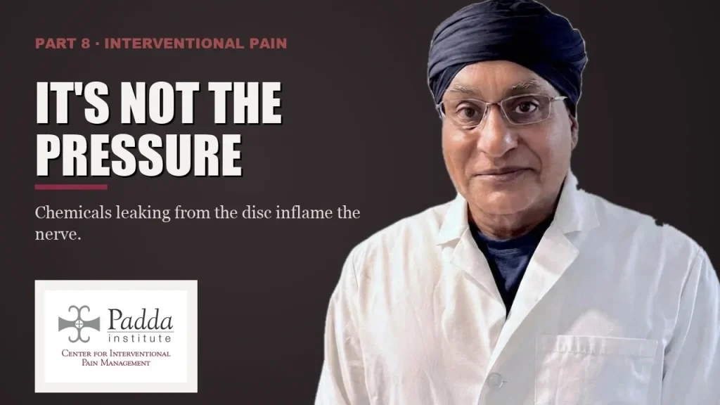
Burning leg or arm pain from a herniated disc is mostly chemical. Escaped disc material bathes the nerve root and its ganglion (a cluster of nerve cells) in inflammatory molecules. The inflamed ganglion then fires with no trigger at all. An epidural steroid injection places corticosteroid at that root, confirmed with fluoroscopy and contrast. The goal is to cut the inflammation, swelling and stray firing.
It is two in the morning and you are standing in your own kitchen, because lying down made it worse. The pain is not really in your back. It is a hot wire running from your buttock, past your knee, down the outside of your calf, into your big toe. Coughing sets it off. Sitting — the car, the desk, the couch — is the worst thing you can do. Walking a little actually takes the edge off. Or it is your arm, and the only way you can sleep is with your hand resting on top of your head. That posture has a name: the shoulder abduction relief sign. This article explains why that pain acts the way it does. It explains what an epidural steroid injection is really doing when it helps. And it lays out what the published evidence says it can and cannot deliver.
What causes burning nerve pain from a herniated disc?
At the center of every spinal disc sits the nucleus pulposus — a gel-like core with no blood supply, walled off from the body. Your immune system has almost never met this tissue. That is thought to be part of why its escape sets off such a strong reaction.
When the outer ring of the disc, the annulus, tears, the inner material pushes out into the epidural space. Two separate injuries begin.
The first is mechanical. Displaced material presses on the nerve root. Pressure tends to block the nerve’s signal. That shows up as numbness in one skin zone, a reflex that drops out, or a weak muscle.
The second is chemical. The evidence suggests it is the chemistry, more than the pressure, that causes the burning. Escaped nucleus pulposus exposes the nerve root — and above all the dorsal root ganglion — to phospholipase A2, tumor necrosis factor alpha, and interleukins. The ganglion is the cluster of sensory nerve cells that sits just outside the spinal canal. This chemical attack is chemical radiculitis.
The ganglion is the weak point. It is less protected than the rest of the root. When it is inflamed, it fires on its own: signal with no trigger at all.
This is also why some people have huge herniations on a scan and no symptoms at all. The image shows shape. It does not show chemistry.
What does a pinched nerve root feel like?
Once you know the ganglion can fire on its own, the strangest part of nerve root pain stops being strange.
Your leg screams while you are lying perfectly still. Nothing presses harder at 2 a.m. than it did at noon. An inflamed ganglion needs no mechanical trigger to fire. So the pain does not need you to be doing anything.
The rest of the pattern is easy to spot. Coughing sets it off. Sitting — the car, the desk, the couch — is often the worst thing you can do. Standing up and walking a little takes the edge off. In the arm, the only way to sleep is with a hand resting on top of the head: the shoulder abduction relief sign. None of these positions prove anything on their own, and the reviews cited below do not test them. What they do is point at a nerve root, not a muscle. That is where the workup has to start.
Numbness and weakness feel like a different problem than the burning. In a sense they are. Numbness, a lost reflex, and weakness track the mechanical injury. The burning tracks the chemical one. That split matters. A scan can show the mechanical injury. It cannot show the chemical one.
What the standard approach often misses
Two failures are common, and neither is subtle.
The series of three, booked before the diagnosis. Patients are handed three epidural steroid injections, six weeks apart and booked in advance. This happens before anyone has decided which nerve root is causing the pain. The number three is not a finding of biology. It is a scheduling habit. A treatment plan that exists before a diagnosis is not a treatment plan.
Numbers borrowed from a different condition. Say your imaging shows lumbar spinal stenosis — and your legs go heavy and dead after two blocks of walking, then recover the moment you sit or lean on a cart. You may be quoted success figures that came from patients with disc herniation and true nerve root pain. The 2025 American Academy of Neurology systematic review looked at lumbar spinal stenosis. It found that epidural steroid injections did not significantly reduce short-term pain (success rate difference −3.5%, 95% CI −12.6 to 5.6). There was possible improvement in disability (short-term success rate difference −26.2%, 95% CI −52.4 to 3.6 — a range that still includes no benefit at all). That is a truly different promise from pain relief. For cervical spinal stenosis, the same review found too little evidence to judge. An earlier meta-analysis in Annals of Internal Medicine also found no clear effect of epidural corticosteroid injections for spinal stenosis.
There is also a technical failure worth naming. An injection done without live imaging, or with imaging but no contrast, cannot be proven to have reached its target. That is exactly why the FDA-convened safety group of many specialties put image guidance near the center of its advice.
What can an epidural steroid injection do, and what are the risks?
Where the medication goes. A transforaminal injection places medicine in the front of the epidural space, right onto the sleeve of the inflamed root. That is where the chemistry is happening. An interlaminar approach covers a wider area at the back. A caudal approach suits anatomy changed by surgery. The full procedure details, including how to prepare and recover, are on the epidural steroid injection treatment page.
What the steroid does once it is there is less settled than you may have been told. Corticosteroid is thought to calm the inflammation at the root — but that mechanism comes from animal studies, not proof in patients. We know the injection helps some people for a while. We do not fully know why. Any clinician who tells you otherwise is overstating the science.
The numbers, plainly. Armon and colleagues, writing in Neurology in 2025 for the American Academy of Neurology, reviewed ninety randomized trials. For cervical and lumbar radicular pain (pain from a nerve root), epidural steroid injections probably reduce short-term pain: a success rate difference of 24.0% (95% CI 12.6% to 34.9%), number needed to treat 4. The authors said plainly that the outcome measures varied too much to give one combined effect size. So no sharper number exists. For longer-term pain relief, the same review found too little evidence. Chou and colleagues, in Annals of Internal Medicine in 2015, found quick improvement averaging 7.55 points on a 100-point scale (95% CI 3.74 to 11.4). That is small, and it did not last.
“Short term” means three months or less in these reviews. Repeat injections are limited by total steroid dose over time. What an injection buys is a window — the stretch of time when rehab becomes possible. It is not a cure.
What you are consenting to. Expect a rise in blood sugar for several days, flushing, insomnia, and possibly a pain flare in the first 48 hours. The needle can pierce the dura (the sac around the spinal nerves) and leave a headache that changes with position and lasts days. Any needle through skin carries a real, if small, risk of infection. Catastrophic neurologic injury, including stroke and spinal cord injury, is rare — but it has happened. Rare is not zero. That is why Rathmell and colleagues published seventeen clinical considerations in Anesthesiology in 2015 for preventing neurologic complications. It is why particulate steroid is not used for cervical transforaminal injections. And it is why a nonparticulate steroid such as dexamethasone is advised for a first lumbar transforaminal injection.
How do you know if an epidural steroid injection is right for you?
First, prove it is a nerve root at all. In the leg, other problems mimic nerve root pain. Hip osteoarthritis causes groin pain, worse when the hip turns inward. Greater trochanteric pain syndrome makes lying on that side impossible. Facet and sacroiliac pain usually stops above the knee. Diabetic lumbosacral radiculoplexus neuropathy is another mimic. So is polyneuropathy, which hits both sides evenly in a stocking pattern, not along one nerve’s map. In the arm: rotator cuff disease, cubital tunnel, carpal tunnel, and thoracic outlet compression.
Resolution requires concordance. Pattern, exam, sensory and reflex findings, and imaging should all point at one level. Nerve testing is added when the picture stays muddy.
When two adjacent levels are both plausible, a selective nerve root block becomes a test, not a treatment. Numbing medicine is placed on one root, and how you feel over the next few hours is the readout. Its honest limit: the numbing medicine spreads past the level it was aimed at, and the evidence for the test is modest. It is used to break a tie, not to invent certainty. For the same reason, a test block should not be done under deep sedation. You need to be awake enough to report what you feel.
Every injection under live fluoroscopy with contrast. Contrast should outline the root before any steroid goes in. If contrast enters a blood vessel, the needle moves. Digital subtraction is used to check the vessels.
Metabolic status is part of the plan, not a substitute for it. If you are insulin resistant, the steroid will push your glucose higher and keep it up longer. So that is managed as part of the procedure. Nothing on a nutrition plan will take pressure off a root or clear a herniation. Omega-3-derived resolution mediators and near-infrared photobiomodulation remain adjunctive and still emerging — they are not why a nerve root calms down.
What a realistic course looks like. What follows is a composite — a picture drawn from many patients with this condition, not one person’s chart, and not a trial result. Someone comes in after eleven weeks of pain running into the sole and outer edge of the foot. The ankle reflex is weak. An MRI shows a disc extrusion at L5-S1 touching the S1 root. Everything lines up, so a transforaminal injection is done at S1 under live fluoroscopy with contrast. The first two days are worse, not better. That flare is expected, and still frightening at three in the morning. Blood sugar runs high for four days. By day ten the pain has dropped enough to sleep flat. That is when the real work starts: nerve gliding, hip and trunk strengthening, and rebuilding the confidence to bend. Around the third month some leg pain creeps back, though not to where it began, and a second injection is done. Function holds better than pain does. That pattern is common; it is not what every patient experiences. Individual results vary.
Ask which root. Ask to see the contrast. Insist on a diagnosis before a series.
Frequently asked questions
Why does my leg burn when I am lying completely still?
Because the pain is chemical as well as mechanical. Inflammatory molecules from disc material irritate the dorsal root ganglion. An inflamed ganglion fires on its own — it makes signal with no trigger at all. That is why changing position does not reliably switch the burning off. It is also why the burning can be severe even when the herniation on your MRI looks small. Individual results vary.
For the other structures besides a disc that can generate leg pain, read Sciatica Is Not a Diagnosis: The Three Structures That Actually Generate Leg Pain.
Do I really need three epidural steroid injections?
No — a pre-booked series of three is a scheduling habit, not a finding of biology. Whether to repeat should follow how you actually responded to the first injection. Repeat injections are limited by total steroid dose over time. What matters more than the number is that the level was found first, with pattern, exam, and imaging all lining up.
For how often steroid injections can be repeated, read How Often Can You Have a Cortisone or Steroid Injection?
How long does the relief last?
In the published reviews, “short term” means three months or less. The benefit is described as small and not lasting. For longer-term pain relief, the 2025 American Academy of Neurology review found too little evidence. Think of an epidural steroid injection as a window in which rehab becomes possible, not as a cure. Individual results vary.
Will an epidural steroid injection help my spinal stenosis?
The evidence here is truly weaker, and you should be told so. In lumbar spinal stenosis, the 2025 American Academy of Neurology review found no significant short-term pain benefit. It found only possible improvement in disability — and the range around that estimate still includes no benefit. For cervical spinal stenosis, the evidence was too thin to judge. If your imaging says stenosis, the success figures for nerve root pain do not apply to you.
For how stenosis causes leg pain that comes on with walking, read Leg Pain When You Walk That Eases When You Lean Forward.
What side effects should I expect, and does it matter that I am diabetic?
Expect a rise in blood sugar for several days, flushing, insomnia, and possibly a pain flare in the first 48 hours. If you are insulin resistant, the steroid will push your glucose higher and keep it up longer. So glucose control is planned as part of the procedure, not left to chance. Less often, the needle can pierce the dura and leave a headache that changes with position and lasts days. Any needle through skin carries a small risk of infection. Catastrophic neurologic injury, including stroke and spinal cord injury, is rare but has happened. That is why a group of many specialties, convened with the FDA, published seventeen safety considerations, including image guidance and nonparticulate steroid. Do not start, stop, or change any medication without consulting your physician.
Where is this done, and how do I get evaluated?
Padda Institute Center for Interventional Pain Management, 4477 Woodson Rd, Suite 100, St. Louis, MO 63134 — right next to St. Louis Lambert International Airport — with a second location at 12174 Natural Bridge Rd, St. Louis, MO 63044. We serve the St. Louis region, Missouri and Illinois. Call (314) 481-5000 or text (314) 886-5902, Monday through Friday, 8:00 a.m. to 5:00 p.m. Bring your imaging and your history.
Our Locations page has the address, hours and directions.
What is the success rate of an epidural steroid injection?
It depends on the condition. The 2025 American Academy of Neurology review covered ninety randomized trials. For cervical and lumbar nerve root pain, it found epidural steroid injections probably reduce short-term pain. The success rate difference was 24.0%, and the number needed to treat was 4. Short term means three months or less. In lumbar spinal stenosis, the same review found no significant short-term pain benefit. Individual results vary.
What happens during an epidural steroid injection?
Every injection is done under live fluoroscopy with contrast. The contrast should outline the nerve root before any steroid is given. If it enters a blood vessel, the needle is moved, and digital subtraction is used to check the vessels. The approach depends on the target. Transforaminal goes right onto the inflamed root. Interlaminar covers a wider area. Caudal is used when surgery has changed the anatomy.
What is the next step if an epidural steroid injection doesn’t work?
Go back to the diagnosis. Leg pain that looks like a pinched nerve can come from hip arthritis, greater trochanteric pain syndrome, the facet or sacroiliac joints, or a nerve disease. Pattern, exam, reflexes and imaging should all point to one level. Nerve testing is added when the picture stays muddy. When two levels side by side are both possible, a selective nerve root block can break the tie.
Key takeaways
- Nerve root pain has two injuries in it. Mechanical pressure causes numbness, reflex loss, and weakness. Chemical inflammation at the dorsal root ganglion causes the burning.
- For cervical and lumbar nerve root pain, a 2025 review of ninety randomized trials found epidural steroid injections probably reduce short-term pain — a 24.0% success rate difference (95% CI 12.6% to 34.9%), NNT 4. The outcome measures varied too much to give one combined effect size. There was too little evidence on longer-term pain relief.
- Those figures do not apply to spinal stenosis. In lumbar stenosis the same review found no significant short-term pain benefit, only possible disability improvement. In cervical stenosis the evidence was too thin.
- The injection buys a window for rehab, not a cure. Here, “short term” means three months or less. Repeat injections are limited by total steroid dose over time.
- Insist on a diagnosis before a series. Know which root, confirmed by pattern, exam, and imaging that all line up. Get it injected under live fluoroscopy with contrast.
This article is educational and is not a substitute for evaluation, diagnosis, or treatment by a physician. Individual results vary. Do not start, stop, or change any medication without consulting your physician. To be evaluated at Padda Institute Center for Interventional Pain Management, call (314) 481-5000 or text (314) 886-5902.
References
- Armon C, Narayanaswami P, Potrebic S, Gronseth G, Bačkonja MM, et al. Epidural Steroids for Cervical and Lumbar Radicular Pain and Spinal Stenosis Systematic Review Summary: Report of the AAN Guidelines Subcommittee. Neurology. 2025;104(5):e213361. PMID 39938000. PubMed
- Chou R, Hashimoto R, Friedly J, Fu R, Bougatsos C, Dana T, Sullivan SD, Jarvik J. Epidural Corticosteroid Injections for Radiculopathy and Spinal Stenosis: A Systematic Review and Meta-analysis. Ann Intern Med. 2015;163(5):373-381. PMID 26302454. PubMed
- Rathmell JP, Benzon HT, Dreyfuss P, Huntoon M, Wallace M, et al. Safeguards to prevent neurologic complications after epidural steroid injections: consensus opinions from a multidisciplinary working group and national organizations. Anesthesiology. 2015;122(5):974-984. PMID 25668411. PubMed
Get the diagnosis before you accept the procedure
Bring your imaging and your history to the Padda Institute Center for Interventional Pain Management in St. Louis. We will tell you which structure is actually generating your pain — and what the evidence does and does not support.
Or call or text us.
Medically reviewed by Dr. Gurpreet Singh Padda, MD, MBA, MHP — Board Certified in Anesthesiology, Pain Medicine, Interventional Pain Management, Addiction Medicine, and Obesity Medicine. Last reviewed July 2026.
Dr. Gurpreet Singh Padda, MD, MBA, MHP