


Main Banner

Ultrasound-Guided and Fluoroscopic Procedures in St. Louis

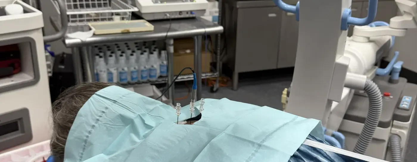
Every injection we perform is placed under image guidance. Which imaging we use depends on the target, and the choice changes what the procedure can tell us.

Why image guidance is not optional

Landmark-based injection, placing a needle by feel and anatomy alone, misses its target more often than most patients realize. When an injection is both a treatment and a diagnostic test, a missed target does not simply fail to help; it produces a false answer that sends the whole workup in the wrong direction. Guidance is what makes the result trustworthy.

What ultrasound adds

Ultrasound shows soft tissue in real time and without radiation. That means a muscle, ligament, tendon or joint can be examined while it moves, under load, in the position that actually reproduces the pain. A tendon that looks intact at rest may show its problem only when the joint is taken through range. This dynamic assessment produces an immediate answer in the room, and the needle, the nerve and the spreading fluid are all visible as the procedure is performed. It is essential for hydrodissection and for peripheral nerve stimulation.

What fluoroscopy adds

Fluoroscopy is live X-ray. It shows bone, and with contrast it shows exactly where injected fluid travels. For spinal work this is the standard: contrast confirms the needle is in the epidural space, in the facet joint, or at the correct nerve root before any medication is given, and it reveals when scarring is preventing normal spread. Epidural steroid injections, facet joint injections and discography all depend on it.

Choosing between them

The target decides. Soft tissue, peripheral nerves and superficial joints favor ultrasound. Deep spinal structures, bone and anything requiring contrast spread favor fluoroscopy. Some procedures use both. Because we have both available on site, the imaging is chosen to suit the problem rather than the problem being forced to fit the available equipment.

What the accuracy data shows

The case for image guidance is not theoretical. Studies comparing landmark-based injection with image-guided injection consistently find that a meaningful proportion of blind injections do not reach the intended target, with accuracy varying by joint and by the experience of the operator. The knee is relatively forgiving; the hip, the shoulder and the small joints of the spine are not.

The consequence is worse than a wasted appointment. When an injection is also a diagnostic test, a missed target produces a false negative. The patient is recorded as not having responded, the true pain source is excluded from consideration, and the workup moves on in the wrong direction. Image guidance is therefore as much about protecting the validity of the diagnosis as about delivering the drug.

Two modalities, different physics

Ultrasound and fluoroscopy answer different questions because they interact with tissue differently. Ultrasound images acoustic impedance differences, so it resolves soft tissue, nerves, tendons and vessels, and it does so in real time with no ionizing radiation. That combination is what permits dynamic assessment: a tendon can be examined while the joint moves through the range that reproduces the symptom, which no static image can replicate.

Fluoroscopy transmits X-rays, so it images bone, and with radiopaque contrast it shows exactly where injected fluid travels. Nothing in ultrasound provides that. Confirming epidural spread, demonstrating intra-articular placement in a facet joint, or revealing that scar tissue is blocking distribution all require seeing the contrast pattern. Radiation dose is managed with pulsed fluoroscopy and collimation rather than by avoiding the modality, because for spinal work the confirmation it provides has no substitute.

Frequently Asked Questions

Ultrasound uses no radiation at all. Fluoroscopy uses short, pulsed exposures, and using it well typically reduces total procedure time and the number of needle passes required.

Yes. With ultrasound in particular, patients frequently watch the screen during the examination, and we will show you what we are seeing.

Marginally, if at all. Accurate first-pass placement usually offsets the setup time, and it substantially improves the reliability of the result.

Request an Appointment

Same-day and emergency appointments are available. Call (314) 481-5000 or request a time below.

Related Treatments and Conditions

Other procedures and conditions our St. Louis pain specialists treat.
Electrodiagnostics

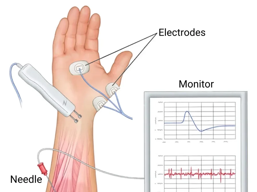
Electrodiagnostics (EDX) testing is used to evaluate the integrity and function of the peripheral nervous (most cranial nerves..

Hip Joint Injection

The hip joint is a large joint where the leg joins the pelvis, functioning as a ball and socket joint. The top of the thigh bone (the femur)…

Discogram

The disks are soft, cushion-like pads which separate the hard vertebral bones of your spine. A disk may be painful when it bulges, herniates..