What this video covers
- Why disc degeneration, bulges, and annular fissures show up in a high share of pain-free people, and what that means for your report.
- What MRI is truly best at: ruling out tumor, infection, fracture, and cauda equina compression, and grading how badly a nerve root is pinched.
- What fluoroscopy adds: live X-ray, real-time contrast, and exact needle placement into deep spinal targets, and when CT guidance is preferred instead.
- What ultrasound adds: real-time images of nerves, tendons, vessels, and outer joints, with no radiation.
- How a diagnostic anesthetic block tests an idea instead of just treating a symptom, and why confirmatory blocks are used.
- The honest limits of each method, including false-positive rates, radiation exposure, ultrasound’s inability to see through bone, and the real but uncommon risks of the blocks themselves.
- MEDICAL DISCLAIMER: This content is for educational purposes only and is not medical advice. It does not substitute for professional diagnosis or treatment. Always consult a licensed healthcare provider regarding your condition. Viewing this video does not establish a doctor-patient relationship.
An MRI report describes tissue, not pain. Disc degeneration, bulges and annular fissures turn up in a high share of people with no pain at all. MRI rules out tumor, infection, fracture and nerve compression. Fluoroscopy and ultrasound guide the needle. A diagnostic anesthetic block tests whether a suspected structure is the pain source.
You are holding a report. Degenerative disc disease at L4-L5. Disc bulge. Annular fissure. Someone said “wear and tear,” handed you a referral, and you have carried that page ever since as if it were a diagnosis. It is not. It is the description of a photograph.
That difference is not a technicality. It is not a criticism of the radiologist who wrote the report — the report did exactly what it was asked to do. A report describes tissue. It does not identify a pain generator: the specific structure whose signals are making the pain you actually feel. This article explains how MRI, fluoroscopy and ultrasound each build their picture. It explains why a picture cannot name the source of pain, and what kind of test can.
How does an MRI make a picture of your spine?
Magnetic resonance imaging does not photograph pain. It listens to hydrogen nuclei — protons — in your tissue. The scanner puts them in a strong magnetic field and tips them with a radio pulse. Then the protons relax back into line. Protons in different chemical settings relax at different speeds. The sequence that is run decides when the scanner listens. That choice is the pulse sequence. It decides which of those differences in relaxing shows up as brightness on the screen.
That is why the same disc can look dark on one sequence and bright on the next. An MRI is not one picture of your spine. It is a set of weighted maps. Each one is tuned to show a different property of the tissue.
That map has no substitute for one job in particular: exclusion. It is how a physician rules out tumor, discitis or epidural abscess (spine infections), hidden fracture, and cauda equina compression. It is also how a physician grades how badly a nerve root is pinched. If your history raises any of those, you are imaged right away. No waiting. No trial of conservative care first.
Red flags that override everything else on this page
Learn these features. If any of them describe you, this article does not apply to you. Get evaluated and imaged now, not next month:
- New loss of bowel or bladder control.
- Saddle numbness — the area that would touch a bicycle seat.
- Leg weakness that is getting worse week over week.
- Fever with back pain.
- Unexplained weight loss.
- Any history of cancer.
- Intravenous drug use.
- Major trauma, or even minor trauma if you have osteoporosis or take steroids long term.
Everything that follows is safe advice only because that list exists.
Why doesn’t my MRI report match my pain?
Here is the part almost nobody is given time to explain. The findings printed on most spine reports are very common in people who feel nothing at all.
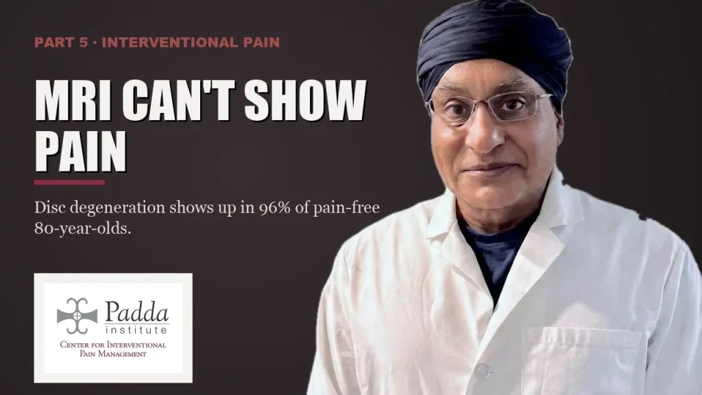
In 2015, in the American Journal of Neuroradiology, Brinjikji and colleagues reviewed 33 studies covering 3,110 people with no back pain. On imaging, disc degeneration appeared in 37% of asymptomatic 20-year-olds and 96% of asymptomatic 80-year-olds. Disc bulges ran from 30% to 84% across the age range, and annular fissures from 19% to 29%. The authors concluded that these findings are likely part of normal aging and unassociated with pain.<sup>1</sup> That review shows how often these features appear in people who were not hurting. It measures prevalence, not causation. That is exactly why it cannot be read as a verdict on your own scan in either direction.
So the line on your report, by the numbers, describes a spine that has been alive for a while. It may also happen to be the source of your pain. The report cannot tell you which, because anatomy is not physiology. A picture shows structure. Pain is a process. A structure is to blame only when silencing it changes your pain.
That is also why scanning first does not reliably help. In 2009, in The Lancet, Chou and colleagues pooled six randomized trials — 1,804 patients. They compared imaging the low back right away against usual care in people without red-flag features. They recorded no significant difference in pain and none in function, at short-term or long-term follow-up. They concluded that imaging without signs of a serious underlying condition does not improve outcomes.<sup>2</sup> Scanning first did not, on average, make people better.
What is the difference between MRI, fluoroscopy and ultrasound?
Each type of imaging answers a question the others cannot.
MRI is a still map of anatomy with excellent soft-tissue contrast. Best question for it: is there something dangerous here, and how compressed is that nerve root?
Fluoroscopy is live X-ray. A pulsed beam passes through you onto a detector. The physician sees your skeleton in real time at any angle the C-arm is turned to. Injected contrast can be watched as it spreads along the target. Or it can be caught running where it should not, into a vessel or toward the intrathecal space (the spinal fluid). That real-time check is why fluoroscopy is the usual choice for needles aimed at deep spinal targets. CT guidance is preferred for some targets with hard anatomy. Best question for it: is this needle, right now, exactly where I intend it to be?
Ultrasound is sound, not radiation. It shows what fluoroscopy cannot see at all: the nerve and its bundles, the tendon, the artery lying beside it, all moving in real time. Its hard limit is physics. Sound does not pass through bone, so deep spinal targets behind bone are not its territory. Best question for it: where exactly is this peripheral nerve, tendon or vessel, and can I place a needle around it without radiation?
None of these beats the others in general. In 2023, in Diagnostics, Viderman and colleagues meta-analyzed eight randomized trials. They compared ultrasound-guided against fluoroscopy-guided procedures for back pain. For pain relief there was no significant difference between the two. For complications there was also no significant difference. But that estimate was too imprecise to rule out a real difference in either direction. Disability scores leaned toward fluoroscopy at the very edge of statistical significance, by a margin too small to matter clinically.<sup>3</sup> Read honestly, those are non-significant findings. On complications, the imprecision means the two cannot be declared equivalent. Non-significant is not the same as proven equal. Pick the method by the target and your anatomy, not by fashion or by what equipment is on hand. You can read more about how ultrasound-guided and fluoroscopic procedures are used for specific targets.
What the usual approach misses
The common path looks like this. Pain, then a scan, then a report full of findings. Then a series of injections aimed at something nobody ever named to you. And no stated bar for what would count as a positive result.
Those injections were not always failed treatments. They were experiments that were never designed, so they could not answer either way. If relief was not defined in advance, and the target was not stated in advance, then relief or no relief tells you nothing about where your pain comes from.
Medication for symptom control has a real and fair place alongside this work. What it does not do is find which structure is making the signal. And a diagnosis put off long enough tends to become a diagnosis abandoned. Do not start, stop, or change any medication without consulting your physician.
How do you find the real source of back pain?
The first move is not the scanner. It is an exam that names a suspect.
Pain in a band along the spine, worse when you lean back and twist, points toward the zygapophyseal (facet) joints and their nerve supply. Pain that follows a nerve path below the knee, with a positive nerve tension sign, points toward the nerve root and the opening it exits through. Pain over the posterior superior iliac spine when standing on one leg raises the sacroiliac joint. Pain on the outside of the hip that comes back when pressed points to the gluteal tendons, not the spine at all.
Now hear how weak those clues are on their own. Dr. Padda covers this in the video above. Published clinical criteria for facet pain were tested against confirmatory blocks in a series of 176 patients. None of the clinical features tested was associated with who responded to the block. No exam move proves your pain comes from the facets. Every finding above is a suspect. None is a verdict.
The verdict needs a test. A diagnostic block is an experiment, not a treatment. A precise amount of local anesthetic goes on one structure. Then we measure what happens to the pain you came in with, recorded in a timed diary.
The anatomy has to be exact
From L1 through L4, the medial branches of the dorsal rami cross the corner where the superior articular process meets the transverse process. That bony corner is the target — not the joint named on your MRI. L5 is the exception that matters most. At L5-S1 there is no medial branch to block. So the L5 dorsal ramus itself is targeted where it crosses the sacral ala and the S1 superior articular process. Get that wrong and you have numbed the wrong nerve and drawn a confident conclusion from it.
One positive block is a hypothesis, not an answer
Diagnostic blocks carry a false-positive rate. The honest move is to give the number, not an adjective. Dr. Padda reports it in the video above. In a published series of 176 patients who had lumbar zygapophyseal blocks, 47% responded to the first block and 15% responded to the confirmatory one. The false-positive rate of a single uncontrolled block was 38%. Nearly two in five of the apparent responses did not hold up.
Because of that, a confirmatory block on a separate day is standard practice at the Padda Institute Center for Interventional Pain Management. That is a practice policy, not a trial result. It also means a large share of first blocks are not confirmed by the second. When that happens, the honest reading is that the wrong structure was tested. So the process goes back to the exam. Individual results vary.
What a block actually buys you
This is the mistake that hurts patients more than the needle does. The relief from a diagnostic block lasts only as long as the anesthetic — roughly one to two hours for lidocaine, several hours for bupivacaine. When it wears off on schedule, that is the test working, not failing. Its whole value is the diary you keep during those hours, even on the days that feel terrible.
Diagnostic blocks are invasive procedures. Bleeding, infection, short-lived nerve irritation, fainting reactions, and reactions to contrast or anesthetic are uncommon but real. Risk rises if you take blood thinners or if the target sits near the spinal cord. Fluoroscopy uses ionizing radiation, kept low with pulsed imaging and a tightly narrowed beam. And blocks are not a screening test. If pain is widespread with no clear target, a needle will not invent one.
An illustrative composite — not a patient testimonial
What follows is a composite built from many patients with this picture, not one person’s chart. Two years of one-sided low back pain, no leg symptoms. A report listing degeneration at several levels and a bulge. Two epidural injections done elsewhere that were not aimed at what the exam pointed to. The pain sits along the spine, gets worse when leaning back and twisting, and does not travel below the knee. The MRI, read for what it is truly good at, is reassuring: no tumor, no infection, no fracture, no root compression. So the idea is tested — a fluoroscopically guided medial branch block at the suspected levels, contrast confirmed, diary in hand for the anesthetic window. The first block gives matching relief. A second, on a separate day, agrees. Individual results vary.
That outcome was chosen for the story, and it is not what most first attempts return. It took weeks, not one visit. And the answer was not a cure. It was a location — a structure that had actually been tested. Not a finding that a large share of pain-free people the same age also carry.
Frequently asked questions
Does an MRI show where my pain is coming from?
No. An MRI shows tissue structure. It is excellent at ruling out tumor, infection, fracture and cauda equina compression, and at grading how badly a nerve root is pinched. It cannot show which structure is making the pain, because a picture records anatomy, while pain is a process in the body. A structure is to blame only when numbing it changes the pain you came in with.
My report says degenerative disc disease and a disc bulge. Is that why my back hurts?
Possibly, but the report alone cannot prove it. A review of 33 studies covered 3,110 people with no back pain at all. Disc degeneration appeared in 37% of asymptomatic 20-year-olds and 96% of asymptomatic 80-year-olds, with bulges in 30% to 84%.¹ Those findings are common features of normal aging. Whether yours is also your pain generator has to be tested, not assumed.
Is ultrasound guidance as good as fluoroscopy for injections?
It depends on the target. A meta-analysis of eight randomized trials of image-guided procedures for back pain found no significant difference between ultrasound and fluoroscopic guidance in pain relief. It found none in complications either. But the complication estimate was too imprecise to rule out a real difference, so the two should not be called equivalent.³ Ultrasound cannot see through bone. So deep spinal targets are usually reached under fluoroscopy, with CT guidance for some hard anatomy. The target and your anatomy decide, not the equipment.
My block wore off after a couple of hours. Did it fail?
Almost certainly not. A diagnostic block is built to last only as long as the local anesthetic — roughly one to two hours for lidocaine, several hours for bupivacaine. Relief that ends on schedule is the expected result. What matters is whether the pain you came in with dropped during that window. That is why the timed diary is the real output of the test. Individual results vary.
I take a blood thinner. Can I still have a diagnostic block?
That is a decision for your physician. It depends on the medication, why you take it, and how close the target sits to the spinal cord. Blood thinners raise bleeding risk for these procedures, so they must be reviewed in advance. Do not start, stop, or change any medication without consulting your physician.
What should I bring and ask at an evaluation?
Bring the images themselves, not only the report, along with the dates of any injections you have already had and what happened to your pain afterward. Then ask two questions: which structure is being targeted, and what result would prove that hypothesis wrong. The Padda Institute Center for Interventional Pain Management is at 4477 Woodson Rd, Suite 100, St. Louis, MO 63134, next to St. Louis Lambert International Airport, with a second location at 12174 Natural Bridge Rd, St. Louis, MO 63044. We serve the St. Louis region, Missouri and Illinois. Call (314) 481-5000 or text (314) 886-5902, Monday through Friday, 8:00 AM to 5:00 PM.
What is fluoroscopy used for in pain treatment?
Fluoroscopy is live X-ray. In pain care it shows your spine in real time, from any angle the C-arm is turned to, while the needle is placed. Injected contrast can be watched as it spreads along the target. It can also be caught running into a vessel or toward the spinal fluid space where it should not go. That real-time check is why it is the usual choice for deep spinal targets.
Is fluoroscopy the same as a CT scan?
No. Fluoroscopy is live X-ray. A pulsed beam passes through you onto a detector, and the physician watches your skeleton and the needle in real time. CT guidance is a separate tool, preferred for some targets where the anatomy is hard. The choice between fluoroscopy, CT and ultrasound depends on the target and your anatomy. It does not depend on which machine happens to be in the room.
What is the main drawback of fluoroscopy?
Two limits. It uses ionizing radiation, which is kept low with pulsed imaging and a tightly narrowed beam. And it cannot show the nerve, the tendon or the artery lying beside a target. Ultrasound shows those without radiation. Ultrasound, in turn, cannot see through bone, so deep spinal targets stay with fluoroscopy. Each tool answers a question the others cannot.
Key takeaways
- An MRI is a set of weighted maps of anatomy. Nothing replaces it for ruling out tumor, infection, fracture and cauda equina compression. It cannot identify which structure is making your pain.
- Disc degeneration, bulges and annular fissures are found in large shares of people with no pain at all — 37% of asymptomatic 20-year-olds up to 96% of asymptomatic 80-year-olds. So a report finding is not by itself a diagnosis.
- MRI, fluoroscopy and ultrasound answer three different questions. Across eight randomized trials, ultrasound and fluoroscopic guidance showed no significant difference in pain relief. The complication comparison was too imprecise to be called equivalent.
- A diagnostic block is an experiment with a set endpoint. In the published series cited in the video, a single uncontrolled block carried a 38% false-positive rate. That is why a confirmatory block on a separate day is used.
- Red-flag symptoms — new bowel or bladder changes, saddle numbness, worsening leg weakness, fever, unexplained weight loss, cancer history — mean you get checked now, not watched and waited on.
This article is educational and is not a substitute for evaluation, diagnosis, or treatment by a physician. Individual results vary. Do not start, stop, or change any medication without consulting your physician. To have your pain generator identified rather than guessed at, call (314) 481-5000 or text (314) 886-5902 to request an appointment.
References
- Brinjikji W, Luetmer PH, Comstock B, Bresnahan BW, Chen LE, Deyo RA, Halabi S, Turner JA, Avins AL, James K, Wald JT, Kallmes DF, Jarvik JG. Systematic literature review of imaging features of spinal degeneration in asymptomatic populations. AJNR Am J Neuroradiol. 2015 Apr;36(4):811-6. PMID 25430861. PubMed
- Chou R, Fu R, Carrino JA, Deyo RA. Imaging strategies for low-back pain: systematic review and meta-analysis. Lancet. 2009 Feb 7;373(9662):463-72. PMID 19200918. PubMed
- Viderman D, Aubakirova M, Aryngazin A, Yessimova D, Kaldybayev D, Tankacheyev R, Abdildin YG. Ultrasound-guided vs. fluoroscopy-guided interventions for back pain management: a systematic review and meta-analysis of randomized controlled trials. Diagnostics (Basel). 2023 Nov 18;13(22):3474. PMID 37998610. PubMed
Get the diagnosis before you accept the procedure
Bring your imaging and your history to the Padda Institute Center for Interventional Pain Management in St. Louis. We will tell you which structure is actually generating your pain — and what the evidence does and does not support.
Or call or text us.
Medically reviewed by Dr. Gurpreet Singh Padda, MD, MBA, MHP — Board Certified in Anesthesiology, Pain Medicine, Interventional Pain Management, Addiction Medicine, and Obesity Medicine. Last reviewed July 2026.
Dr. Gurpreet Singh Padda, MD, MBA, MHP