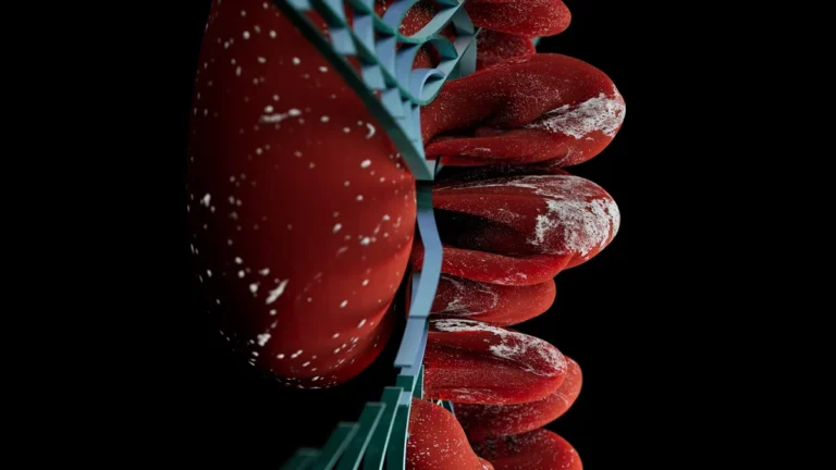
Gut-brain axis inflammation is gut immune trouble reaching the brain by three roads: the vagus nerve carrying inflammatory reports upward, the blood carrying bacterial endotoxin and cytokines through a breached gut wall, and immune cells trained in the gut. Mast cells sit where all three meet. It is measurable neuroinflammation, not stress.
Pooled across 22 studies of 6,344 people with coronary heart disease, the ones who were also depressed carried two signatures at once: more C-reactive protein and interleukin-6 in the blood, and a quieter vagus nerve on the heart tracing, with the beat-to-beat vagal index lower by about 8 milliseconds1. A blood test and a heart monitor do not know the word “stress.” Both found a fire burning and a brake not holding.
Brain fog, low mood, anxiety and pain turned up too loud are what immune traffic from the gut feels like when it arrives. The gut, our first brain, and the brain in your skull, the second, trade that traffic on three roads. The map comes from Chapters 3, 4, 12 and 16 of The Angry Gut.
How does the gut talk to the brain?
Picture a city under siege with three ways out. The first is a phone line: the vagus nerve, which in the abdomen is built almost entirely of slow sensory fiber, so most of its traffic is reports going up. The second is the river: the blood draining the gut, carrying whatever crossed the wall. The third is the army: immune cells and antibodies trained in the gut wall.
Mast cells stand at the gate where all three roads begin. They sit in the gut wall in two-way contact with sensory nerve endings, and they take orders from the lymphocytes and antibodies of the third road. When they fire, histamine, proteases and cytokines excite the nerve and loosen the wall. Part 1 of this series introduces them; how they fire pain nerves is in histamine, mast cells and the nerves of the gut.
Can you measure the gut-brain connection?
You can measure the wire, and the instrument is your heartbeat. Breathe in and the heart speeds up slightly; breathe out and it slows. That flutter is vagal, and a low reading means a brake with worn pads.
In 253 adults studied with 24-hour heart recordings and electrical recordings of the stomach, vagal readings were lowest in people with both severe anxiety-depression and functional dyspepsia. The surprise sits in the group with no stomach complaints at all: in 60 adults with mild to moderate anxiety and depression and no dyspepsia, the stomach’s normal electrical surge after a meal was blunted in every one2. The second brain’s state was already written into the first brain’s response to lunch.
One caveat: heart rate variability reads the vagus at the heart, not the gut. It is a proxy, and I say so every time. Which stimulators beat a sham is in the vagus nerve and pain.
Is gut-brain inflammation real, or is it just stress?
The word “just” is the mistake. Stress is a chemical with targets, and one of them is the gut wall.
In healthy volunteers, a public speech made the small bowel more permeable, but only in people whose cortisol actually rose. Corticotropin-releasing hormone, the brain’s opening order in a stress response, opened the wall by itself, and pretreating with cromolyn, a mast cell stabilizer, blocked both the speech and the hormone3. A later rematch from the same lab was less clean in living volunteers, as what opens the gut wall in an ordinary week explains. The mechanism is real at the tissue and unsettled in the whole person: enough to treat stress as a body input, not a character flaw.
What gets from the gut into the blood?
Now the river. Healthy lean adults ate a 910-calorie fast-food meal after an overnight fast. Within three hours their blood endotoxin, a piece of the outer coat of gut bacteria, rose by about 47%, and their white blood cells switched on TLR4, the endotoxin receptor, and NF-kB, the master switch of inflammation. A second group ate 910 calories of fiber and fruit, and nothing rose, even though that meal carried more measured endotoxin4. The problem is what the meal does to the wall.
Follow the money. The fast-food version is the cheap one a food system built on subsidized calories puts on every corner; the fiber-and-fruit version costs more time and money. That is a supply chain delivering an inflammatory dose three times a day, not a willpower problem.
Does gut inflammation reach the brain?
The brain’s own barrier was never a wall. Contrast MRI in living people shows it leaking with age in one place first: the hippocampus, the memory center. Against young adults, older adults with normal thinking showed 41% more leak in the hippocampus and 107% more in its CA1 region, rising again in mild cognitive impairment, while cortex and white matter did not change5. In type 2 diabetes, white matter leaked before small-vessel disease was visible on a standard scan6. Blood sugar pries that door open before plaque does.
Among 217 adults aged 60 and over, the 33.6% with cognitive impairment carried about three times the interleukin-6, twice the interleukin-1 beta, more LBP (the protein that carries endotoxin) and its receptor TLR4, and about four times the stool calprotectin, a marker of gut inflammation. Their diet quality did not differ7.
This is where I have to be careful with you. That is one snapshot in people whose first brain is already inflamed. It does not prove the endotoxin caused the forgetting, and a resident bacterial colony in the healthy brain has not been shown. What reaches the second brain is traffic across an opening barrier, not a colony. Our position follows: treat the first brain now, because the metabolic reasons to do it stand on their own. How that traffic feels day to day is in Part 3, on brain fog.
Do gut immune cells turn on the brain and nerves?
The third road is the army, and the gut is where it goes to school. In the lymph nodes that drain the gut, specialized cells teach young T cells what to ignore, a mechanism worked out in mice. When the lesson goes wrong, the graduates attack the wrong target.
The cleanest human example starts with food poisoning. Campylobacter jejuni, a gut bacterium, carries surface molecules that resemble parts of the peripheral nerves. Antibodies built against the bug cross-react with the nerves, and the result can be Guillain-Barré syndrome, the most common cause of sudden flaccid paralysis worldwide8. The warning belongs beside it: most microbes proposed as mimicry triggers fail the formal test.
Mast cells sit downstream of this road, because lymphocytes and their antibodies are potent mast cell activators. A miseducated immune system keeps the gatekeepers primed, and primed gatekeepers keep the first two roads busy. That is why our treatment works upstream on the lymphocytes, as Part 7, on MCAS treatment, explains.
How do you calm an inflamed gut-brain axis?
You cannot argue a brain out of inflammation arriving by three roads. You cut the traffic at the source.
- Change the plate, because the wall answers within hours. One fast-food meal raised endotoxin by three hours; the fiber-and-fruit meal did not4. Every meal is a dose you choose.
- Repair the metabolism, because it holds the brain’s barrier open. Diabetes opened the white matter barrier before any damage showed6. Blood sugar and waistline are brain treatment.
- Treat stress as chemistry. The hormone that opens the gut wall through mast cells3 fires whenever the second brain reads threat. Acceptance and Commitment Therapy, delivered in our clinic by a pain-trained behavioral clinician, works on how often it reads threat.
Do this and the wire carries calmer reports, the river runs cleaner, and the army stops drilling for a war that is not coming. The fog, the mood and the pain move last, because they are the end of the road.
Frequently asked questions
Is there a test for brain inflammation coming from the gut?
A single blood test for it does not exist. Heart rate variability shows how well the vagal brake is holding, but it reads the nerve at the heart. In the older-adult study above, the marker that tracked thinking scores most closely was stool calprotectin, a gut inflammation marker, not a blood endotoxin marker7. Fog that nobody has explained deserves a look at the first brain, the same way we approach long-term gut inflammation in chronic pain.
Why does gut inflammation change mood and anxiety?
Cytokines that arrive in the second brain change how it builds its chemical messengers, pulling tryptophan away from serotonin. That turns an immune event into a mood event without any change in your circumstances. The chemistry, and the role histamine plays in it, is laid out in Part 4, on MCAS anxiety.
What is the difference between the gut-brain axis and the enteric nervous system?
The enteric nervous system is the first brain itself: the network of nerve cells built into the gut wall that runs digestion on its own. The gut-brain axis is the traffic between that first brain and the second brain in your skull, carried by the vagus nerve, the blood and the immune system. The first brain’s wiring is described in the enteric nervous system and pain.
The MCAS series, from The Angry Gut: part 5 of 7
- What MCAS is, and why it starts in the gut wall
- MCAS symptoms across gut, skin and brain
- MCAS brain fog
- MCAS and anxiety
- The gut-brain axis and inflammation (you are here)
- The low histamine diet
- MCAS treatment, layer by layer
Want all three roads checked in one visit?
We trace all three roads in one evaluation: the gut wall, the metabolic terrain that holds the brain’s barrier open, and the immune load keeping mast cells primed. One plan, not two offices.
Request an appointment, or call or text us.
Sources
- Li G, Zhang L, Liu M. Meta-analysis on inflammation and autonomic nervous system of coronary heart disease combined with depression. BMJ Open. 2024;14(3):e079980. doi:10.1136/bmjopen-2023-079980 (PMID 38453204)
- Du L, Yang J, Jiang L, Zeng G, Shu Y, Bi B. Postprandial attenuation of gastric slow waves in anxiety-depression, with and without functional dyspepsia: associations with heart rate variability and sleep quality. Frontiers in Medicine. 2026;13:1890716. doi:10.3389/fmed.2026.1890716 (PMID 42577285)
- Vanuytsel T, van Wanrooy S, Vanheel H, Vanormelingen C, Verschueren S, Houben E, et al. Psychological stress and corticotropin-releasing hormone increase intestinal permeability in humans by a mast cell-dependent mechanism. Gut. 2014;63(8):1293-9. doi:10.1136/gutjnl-2013-305690 (PMID 24153250)
- Ghanim H, Abuaysheh S, Sia CL, Korzeniewski K, Chaudhuri A, Fernandez-Real JM, et al. Increase in plasma endotoxin concentrations and the expression of Toll-like receptors and suppressor of cytokine signaling-3 in mononuclear cells after a high-fat, high-carbohydrate meal: implications for insulin resistance. Diabetes Care. 2009;32(12):2281-7. doi:10.2337/dc09-0979 (PMID 19755625)
- Montagne A, Barnes SR, Sweeney MD, Halliday MR, Sagare AP, Zhao Z, et al. Blood-brain barrier breakdown in the aging human hippocampus. Neuron. 2015;85(2):296-302. doi:10.1016/j.neuron.2014.12.032 (PMID 25611508)
- Chen YC, Lu BZ, Shu YC, Sun YT. Spatiotemporal Dynamics of Cerebral Vascular Permeability in Type 2 Diabetes-Related Cerebral Microangiopathy. Frontiers in Endocrinology. 2021;12:805637. doi:10.3389/fendo.2021.805637 (PMID 35087478)
- Arikawa AY, Waterman A, Mishra SP, Labyak C, Williams C, Chaudhari DS, et al. Cognitive Impairment Is Associated with Alterations in Diet Quality and Inflammatory Biomarkers in Older Adults: A Cross-Sectional Analysis of Data Collected from the Microbiome in Aging Gut and Brain (MiaGB) Consortium Cohort. The Journal of Nutrition. 2025. doi:10.1016/j.tjnut.2025.06.026 (PMID 40617511)
- Shahrizaila N, Lehmann HC, Kuwabara S. Guillain-Barré syndrome. Lancet. 2021;397(10280):1214-1228. doi:10.1016/S0140-6736(21)00517-1 (PMID 33647239)
Dr. Gurpreet Singh Padda, MD, MBA, MHP