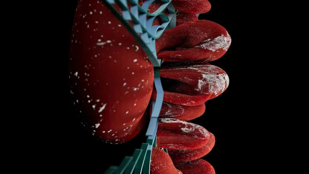
Mast cell activation syndrome (MCAS) is a condition in which mast cells, the immune alarm cells packed into the gut wall, release histamine, proteases and cytokines too easily and too often. Flushing, gut pain, brain fog and anxiety follow. Those cells mostly take their orders from lymphocytes at the gut border, so that is where we look first.
Most people told they have mast cell activation syndrome get handed a drug that quiets a bell. Nobody asks who is ringing it. The answer sits in the lining of your bowel, not in your skin and not in your skull.
In this series the gut is the first brain and the organ in your skull is the second brain. In Chapter 31 of The Angry Gut, which Ami Michelle Grimes and I wrote, the mast cell is not the start of the story. It is the middle. This page is the map.
What is mast cell activation syndrome?
A mast cell is an immune cell packed with granules, like a fire extinguisher that is already pressurized. Fired, it sprays histamine, proteases (enzymes that cut proteins), prostaglandins and cytokines (immune messenger proteins). In MCAS the cells fire too easily, too often, at things that are not a threat.
The textbook calls it an allergy cell. That is wrong. Mast cells sit where the fast, built-in immune system meets the slow, learned one. They take signals from T cells and other immune cells, answer many triggers besides the allergy antibody IgE, and trade signals both ways with sensory nerves1. The mast cell is the alarm. The lymphocytes, and the antibodies they make, hold the switch.
Once released, those chemicals make the gut lining leak and gut nerves more sensitive, and the pain from histamine and proteases runs through prostaglandins made inside the mast cell itself2. The cramping, the loose stool and the leaking wall are one event with three names. The pain side of it is laid out in how gut mast cells fire your pain nerves.
Why does MCAS live mostly in the gut wall?
You have probably read that 70% of your immune system lives in your gut. It was copied from paper to paper, never measured. When researchers counted every immune cell in a reference adult body, the gut held about 3% of them and around 5% of the lymphocytes3.
So why does the gut matter? It is not where the immune system lives. It is where the immune system writes. Every day a human body makes more IgA, the antibody that guards the lining, than IgG and IgM combined4. Your bowel lining is one layer of cells holding back a sewer of bacteria and food, and the immune system writes against it all day. Lymphocytes and their antibodies are among a mast cell’s strongest switches, so a border that writes that much keeps its alarms primed.
Here is the part almost nobody tells you. Gut mast cells also work in the serotonin factory: enterochromaffin cells and mast cells in the lining make about 90% of the serotonin produced in the intestine5. MCAS was never only an allergy problem.
How is MCAS diagnosed?
Two frameworks compete, and they produce very different numbers of patients. Anyone who says MCAS should say which one.
The strict consensus criteria require all three, not any one: symptoms that recur in more than one organ system, a measured rise in a mast cell chemical during an episode, and a response to drugs aimed at mast cells or their chemicals. Normal serum tryptase runs from 0 to 11.4 ng/mL, and an episode counts only if tryptase climbs to at least baseline times 1.2, plus 2 ng/mL6. If your baseline is 5, an attack has to reach 8.
The broad framework argues those rules miss most patients. Its authors put MCAS at up to 17% of the general population, and in the same paper concede that their criteria have the potential to overdiagnose7. That is why I name the ruler before I name the disease. When someone meets only the broad criteria, we treat the same terrain anyway: the gut wall and the lymphocytes behind it.
Are you born with MCAS or does it develop?
Some of it is inherited, and that part can be measured. Hereditary alpha-tryptasemia, an inherited trait in the tryptase gene, turned up in 4% of healthy blood donors and in 29% of people with non-clonal mast cell activation, and carriers had higher baseline tryptase8. A gene can raise the floor. It does not write the attacks.
Most people in that non-clonal group did not carry the trait. Their mast cells are being told to fire by lymphocytes trained, for years, on whatever crossed the gut wall. One smoke detector leaves the factory too sensitive. Another works fine but hangs over the stove. Most MCAS is the second kind, and you can move a stove.
Is MCAS a rare condition, or is everyone getting it?
It depends on the ruler. Strict criteria demand a tryptase rise caught during an attack, and few people qualify. Broad criteria reach up to 17% of the population. Much of the sense that everyone has MCAS is the gap between those rulers.
The rest is terrain: antibody traffic at the wall, and bacterial fragments called endotoxin slipping through a loosened lining to keep the immune cells beneath it on alert. A third driver is not biological at all: in married couples, hostile arguments tracked a blood marker of gut endotoxin leak9. Conflict at home is a gut input, and Part 4 gives the numbers.
How does a gut immune reaction reach the brain?
Serotonin made in the gut cannot cross the barrier around the brain. There is no pipeline from colon to cortex. The first brain reaches the second three other ways.
The wire. Gut serotonin switches on 5-HT3 receptors on vagus nerve fibers, which relay through the brainstem to the amygdala, the brain’s threat center5. It is a phone line, not a pipe; more in the vagus nerve and gut pain signals.
The blood. Cytokines and bacterial fragments from an inflamed wall raise whole-body inflammation, the thing those C-reactive protein numbers measure9.
The chemistry. Inflammation turns on IDO, an enzyme that sends tryptophan, the raw material for serotonin, down a side track toward kynurenine, leaving less to cross into the brain and become serotonin there5. Your brain did not run out of serotonin. Its supply train was rerouted, like freight switched to the wrong town. The same fire feeds worry, as I explain in gut health and anxiety.
Brain fog, anxiety and pain are what that traffic feels like. I will not promise you a number for how much clearer your head gets when the wall closes; that study has not been run. Our position is simpler: the first brain and the second brain are one system, and we treat them as one.
Following the traffic through the next six parts
Part 2, MCAS symptoms explains why the list jumps from skin to bowel to head and keeps growing, and why that is a signal, not a mystery.
Part 3, MCAS brain fog follows the traffic into the second brain: where its barrier opens first, and why metabolic disease opens it more reliably than plaque does.
Part 4, MCAS and anxiety is the immune-made neurotransmitter story in full: why anxiety that rides with a bad bowel is not a chemical imbalance in the head.
Part 5, the gut-brain axis and inflammation is the wiring diagram, separating what neuroinflammation means from what it is sold as.
Part 6, the low-histamine diet asks a blunt question: is the histamine that hurts you the histamine you eat?
Part 7, MCAS treatment covers antihistamines, cromolyn and low-dose naltrexone. Low-dose naltrexone is not a mast cell stabilizer; it works upstream, on the lymphocytes that switch mast cells on, and Part 7 explains how.
What should you do first if you think you have MCAS?
Put every organ on one list. Skin, gut and head, with dates and meals. Split across three specialists, one event gets seen in thirds.
Get the ruler right. Ask for a baseline tryptase when you are well and a second one during an episode. That pair decides which framework you meet.
Feed the wall, not the capsule. What you eat decides which bacteria thrive in the first brain and what they make, histamine included. Changing what you feed the first brain moves the second more reliably than swallowing an organism does.
Take the conflict seriously. If home is a battlefield, your gut lining is keeping score. Our in-house behavioral clinician treats it as protocol, because the endotoxin marker does not care whether stress was chemical or personal.
Quieting the bell has its place, and we use the drugs that do it. Our job is to find the hand on the switch, and in most people with MCAS that hand is at the gut wall.
Frequently asked questions
Is mast cell disease autoimmune?
It sits next door to autoimmunity rather than inside it. In classic autoimmune disease, the immune system aims antibodies or T cells at your own tissue. In most MCAS, the mast cell is answering signals from lymphocytes and antibodies that were trained at the gut border, so the two problems share a source more than a label. The immune system is not confused; it is responding to what it is being shown. That is why our treatment works upstream of the mast cell, as Part 7 on MCAS treatment explains.
Why don’t antihistamines fix MCAS on their own?
Histamine is one weapon out of several. In human gut tissue, the mast cell’s proteases drove more pain-nerve firing than its histamine did, and prostaglandins and cytokines add their own load. An antihistamine blocks one receptor for one chemical, so partial relief is the expected result, not a sign the diagnosis is wrong. The full nerve-level picture is in histamine and pain.
Does leaky gut cause MCAS, or does MCAS cause leaky gut?
The arrow runs both ways, which is what makes it a loop. Mast cell chemicals loosen the gut lining. A looser lining lets bacterial fragments reach the immune cells underneath. Those immune cells keep the mast cells primed, and the cycle turns again. Breaking it means working on the wall and the immune traffic at the same time; how the lining fails is covered in leaky gut symptoms and chronic pain.
Why would a pain practice evaluate mast cell activation?
Because gut mast cells sit against sensory nerves, and their chemicals excite the fibers that carry pain. Belly pain that arrives with flushing, loose stool and brain fog is one problem with one source, and treating the pain alone leaves the source running. That pattern is laid out in Part 2 on MCAS symptoms.
The MCAS series, from The Angry Gut: part 1 of 7
- What MCAS is, and why it starts in the gut wall (you are here)
- MCAS symptoms across gut, skin and brain
- MCAS brain fog
- MCAS and anxiety
- The gut-brain axis and inflammation
- The low histamine diet
- MCAS treatment, layer by layer
Is your gut wall setting off the rest of you?
We evaluate the gut, the immune system and the brain as one system, starting with the tryptase pair that decides which MCAS framework you meet. Bring your whole symptom list, skin to bowel to head.
Request an appointment, or call or text us.
Sources
- Stassen M, Hartmann AK, Delgado SJ, Dehmel S, Braun A. Mast cells within cellular networks. The Journal of Allergy and Clinical Immunology. 2019;144(4S):S46-S54. doi:10.1016/j.jaci.2019.01.031 (PMID 30731122)
- Hasler WL, Grabauskas G, Singh P, Owyang C. Mast cell mediation of visceral sensation and permeability in irritable bowel syndrome. Neurogastroenterology and Motility. 2022;34(7):e14339. doi:10.1111/nmo.14339 (PMID 35315179)
- Sender R, Weiss Y, Navon Y, Milo I, Azulay N, Keren L, et al. The total mass, number, and distribution of immune cells in the human body. Proceedings of the National Academy of Sciences of the United States of America. 2023;120(44):e2308511120. doi:10.1073/pnas.2308511120 (PMID 37871201)
- Conley ME, Delacroix DL. Intravascular and mucosal immunoglobulin A: two separate but related systems of immune defense?. Annals of Internal Medicine. 1987;106(6):892-9. doi:10.7326/0003-4819-106-6-892 (PMID 3579073)
- Hwang YK, Oh JS. Interaction of the Vagus Nerve and Serotonin in the Gut-Brain Axis. International Journal of Molecular Sciences. 2025;26(3):1160. doi:10.3390/ijms26031160 (PMID 39940928)
- Valent P, Akin C, Nedoszytko B, Bonadonna P, Hartmann K, Niedoszytko M, et al. Diagnosis, Classification and Management of Mast Cell Activation Syndromes (MCAS) in the Era of Personalized Medicine. International Journal of Molecular Sciences. 2020;21(23):9030. doi:10.3390/ijms21239030 (PMID 33261124)
- Afrin LB, Ackerley MB, Bluestein LS, Brewer JH, Brook JB, Buchanan AD, et al. Diagnosis of mast cell activation syndrome: a global "consensus-2". Diagnosis. 2021;8(2):137-152. doi:10.1515/dx-2020-0005 (PMID 32324159)
- González-de-Olano D, Navarro-Navarro P, Muñoz-González JI, Sánchez-Muñoz L, Henriques A, de-Andrés-Martín A, et al. Clinical impact of the TPSAB1 genotype in mast cell diseases: A REMA study in a cohort of 959 individuals. Allergy. 2024;79(3):711-723. doi:10.1111/all.15911 (PMID 37818990)
- Kiecolt-Glaser JK, Wilson SJ, Bailey ML, Andridge R, Peng J, Jaremka LM, et al. Marital distress, depression, and a leaky gut: Translocation of bacterial endotoxin as a pathway to inflammation. Psychoneuroendocrinology. 2018;98:52-60. doi:10.1016/j.psyneuen.2018.08.007 (PMID 30098513)
Dr. Gurpreet Singh Padda, MD, MBA, MHP