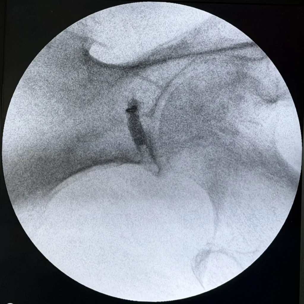
A selective nerve root block tests whether the bulging disc on your MRI is actually causing your leg pain. A small volume of local anesthetic goes to a single nerve root under fluoroscopic guidance (live X-ray). If the leg pain that root carries goes quiet, that root is implicated. If nothing changes, it is not.
A bulging disc is the most common explanation given for sciatica, and it is often the right one. The problem is that it is also the one most likely to be taken on faith, without a test, because a low-back MRI almost always shows something that could plausibly be blamed.
Here is the number that should set how much weight a scan gets. A systematic review looked at imaging in 3,110 asymptomatic people — people with no back pain at all. Disc bulges showed up in 30% of 20-year-olds, rising to 84% of 80-year-olds. Disc degeneration (wear) ran from 37% to 96% across the same ages. Disc protrusion ran from 29% to 43%, and annular fissures (small tears in the disc wall) from 19% to 29%.1 The authors concluded that many wear-and-tear findings on scans are likely normal aging and not tied to pain. They must be read in light of the patient’s actual condition.1
So a report that shows a bulge at L4–5 has told you that you have a common finding. It has not told you that this finding is causing your leg pain.
The test that answers what the scan cannot
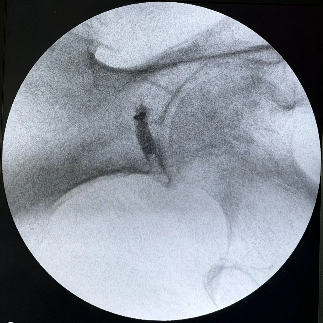
A selective nerve root block is a test first. A small volume of local anesthetic goes to a single nerve root under fluoroscopic guidance. Contrast dye goes in first to show exactly where the fluid sits. If the leg pain that root carries goes quiet, that root is implicated. If nothing changes, it is not.
This is the same logic as a piriformis injection, used one structure over. In both cases the medicine is not the point. The point is whether shutting off one suspect changes your pain.
Why this is not an epidural
The difference is easy to miss, and it matters. An epidural steroid injection is built to spread across a region. That makes it a fair treatment and a poor test. A selective nerve root block is built to stay put. When a block spreads, it stops being selective. Then its result can no longer be trusted.
How accurate is it, honestly?
Moderately accurate, and it is worth giving real numbers instead of comfort. A prospective, controlled, single-blinded study did 105 fluoroscopically guided blocks in 47 patients. Each had pure radiculopathy (pain from an irritated nerve root) from a single confirmed level. 47 blocks were at the painful level and 58 at a nearby pain-free control level. The cutoff was 70% pain relief, set by ROC analysis. The block showed a sensitivity of 57%, specificity of 86%, accuracy of 73%, positive predictive value of 77% and negative predictive value of 71%.2
The authors’ own summary: the accuracy of diagnostic lumbar selective nerve root blocks is only moderate.2 That is more useful to know than a sales pitch. It shapes how to use the result. It is strong evidence, weighed alongside the exam. It is not the single deciding vote.
What causes the wrong answers — and why contrast fixes most of it
The same study found what caused its false results. All of them could be seen on the spot X-rays:2
- False negatives — insufficient infiltration, insufficient passage of the injectate, and intraepineural injection. In plain terms: the numbing medicine never fully reached the root, so a real pain source was missed.
- False positives — overflow of injectate from the pain-free level being tested into the epidural space or into the painful level. The wrong root got the credit because the fluid did not stay where it was put.
Every one of those failures is a spread problem. Every one shows up on the contrast images. That is why a block done without recorded contrast is not just less precise — its result cannot be graded, because there is no way to tell a true negative from an inadequate injection.
When the disc is not the answer
Say a well-performed, contrast-documented block at the level your MRI flagged does not change your leg pain. That is information worth acting on. It argues against the structure everyone had been treating. It shifts attention to other suspects that cause the same symptoms with no pinched nerve root:
- The deep gluteal space — piriformis syndrome, gemelli-obturator internus syndrome, ischiofemoral impingement, proximal hamstring syndrome.
- The hip joint itself, which sends pain into the buttock more often than textbooks suggest.
- The sacroiliac joint.
- The cluneal nerves over the posterior iliac crest.
Each of those can be tested the same way. That is why we build the workup this way. Sciatica names a route down the leg. It does not name what started it.
The sequence that avoids wasted years
The failure pattern is easy to spot. The MRI shows a bulge. Treatment is aimed at the bulge. Treatment does not work. Treatment is repeated with more force. Each step makes sense on its own, but no one goes back to check the first guess.
The answer is not more imaging. It is testing the suspect the scan pointed to. It is recording the test well enough to trust its answer. And it is treating a negative result as progress.
Frequently asked questions
Does a bulging disc on MRI mean it is causing my sciatica?
Not on its own. A systematic review looked at 3,110 people with no symptoms at all. Disc bulges showed up in 30% of 20-year-olds and 84% of 80-year-olds. Disc degeneration showed up in 37% and 96%. Those are pain-free people. A bulge on your scan is a suspect, not a verdict — and the older you are, the weaker the link. Learn more: herniated disc.
What is a selective nerve root block and how is it different from an epidural?
An epidural steroid injection is built to spread — it bathes a region. That is useful for treatment and useless for finding one level. A selective nerve root block does the opposite. A small volume of local anesthetic goes to one nerve root under fluoroscopic guidance. Contrast confirms it stayed there. If your leg pain goes quiet, that root is implicated. Spread is the enemy of the test. Learn more: selective nerve root block.
How accurate is a selective nerve root block?
Moderately, and it is worth knowing the real numbers instead of comfort. A prospective controlled study did 105 blocks in 47 patients with radiculopathy from a single confirmed level. With a 70% pain-relief cutoff, the block had a sensitivity of 57%, specificity of 86%, accuracy of 73%, positive predictive value of 77% and negative predictive value of 71%. Useful, clearly imperfect, and better than deciding from an MRI alone. Learn more: why contrast confirmation matters.
Why does the contrast picture matter during a nerve root block?
Because it explains the wrong answers. In that same study, false negatives traced to insufficient infiltration, insufficient passage of the injectate and intraepineural injection. False positives came from the injectate overflowing out of the pain-free level into the epidural space or into the painful level. Every one of those failures is visible on the contrast images. Without them, a result cannot be graded. Learn more: reading a diagnostic block result.
My MRI shows bulges at two levels. Which one is the problem?
That is exactly the question a selective nerve root block exists to answer. And it is common — wear at many levels is the rule, not the exception, past middle age. We block one level at a time, with contrast showing the fluid stayed put. That is how a two-level or three-level MRI shrinks to one clear target instead of a broad guess. Learn more: when the answer is not a disc at all.
What if the nerve root block does not help either?
Then the disc is probably not your pain source. That is a real result, not a dead end. It shifts attention to the structures that cause the same leg pain without a nerve root — the deep gluteal muscles, the sacroiliac joint, the hip joint and the cluneal nerves. Each can be tested the same way. Learn more: hip arthritis or sciatica.
To discuss your own case, request an appointment through the appointment request form, call (314) 481-5000, or text (314) 886-5902. You can review the full range of pain treatments, read about the pain management doctors in St. Louis, or find both offices on the locations page.
Sources
- Brinjikji W, Luetmer PH, Comstock B, et al. “Systematic literature review of imaging features of spinal degeneration in asymptomatic populations.” AJNR American Journal of Neuroradiology. 2015;36(4):811–816. doi:10.3174/ajnr.A4173
- Yeom JS, Lee JW, Park KW, Chang BS, Lee CK, Buchowski JM, Riew KD. “Value of diagnostic lumbar selective nerve root block: a prospective controlled study.” AJNR American Journal of Neuroradiology. 2008;29(5):1017–1023. doi:10.3174/ajnr.A0955
Dr. Gurpreet Singh Padda, MD, MBA, MHP