When low back pain sits low and to one side, and scans keep coming back normal, the sacroiliac joint is one of the structures worth testing directly. Up to 25% of low back pain may come from the sacroiliac joint. Yet it is easy to miss, because the joint barely moves, and its pain spreads in patterns that mimic sciatica.1 It is not confirmed with a scan. It is confirmed with an image-guided injection that numbs the joint. Then you see if the pain it is supposed to cause actually goes away.
Why the sacroiliac joint is easy to overlook
The sacroiliac joints pass load from the upper body to the legs. Strong ligaments and muscles hold them. They barely move, only about 2 mm to 4 mm in any direction.1 A joint that moves that little does not show much on a film. That is one reason a patient can have serious pain while the scan shows no clear target.
About 85% of people get low back pain at some point in life. In about 25% of those patients, the sacroiliac joint may be the cause.1 That is a large group of people whose pain gets blamed on the low back by default.
The referral patterns are the reason it gets called sciatica
Patients with a sacroiliac joint problem alone often feel pain just below and toward the midline from the bony bump at the back of the pelvis (the posterior superior iliac spine). Pointing one finger at that spot is the Fortin finger test.1 But pain in one spot is not reliable on its own. A 2000 study found 18 different pain patterns from the sacroiliac joint. Pain ran down the back or side of the thigh in 50%, below the knee in 28%, and into the foot in 14%.1
Pain that runs below the knee is widely assumed to be a pinched nerve root. It is not always. That assumption is how a sacroiliac problem ends up being worked up as sciatica from a disc or the piriformis and treated at the wrong level.
What the examination can and cannot establish
A full history and physical exam come first. The exam must consider other sources that send pain to the same area: the mid and low spine, the pelvic organs, and the hip.1 Range of motion, tenderness, and a nerve exam that checks feeling, strength and reflexes all narrow the field.
Provocation tests are part of that exam. In the sacral thrust test, the patient lies face down. The examiner presses down with the heel of the hand on the center of the sacrum (the flat bone at the base of the spine). Pain in the sacroiliac area suggests a problem. No single test settles it. Three or more positive provocation tests make the diagnosis more accurate.1
Imaging rules things out more than it rules things in
Imaging mainly helps rule out other sources. Plain X-rays show hip disease and structural change at the sacroiliac joint. MRI picks up about 90% of spondyloarthritis (an inflammatory spine arthritis). But it is not useful for problems that are not inflammatory. And nuclear bone scans are not a useful tool for finding sacroiliac joint problems.1
That gap between what a scan shows and what is causing the pain is not unique to this joint. It is the same problem described in what imaging can and cannot show about pain and in deep low back pain with a normal MRI.
Why the injection is the diagnostic test
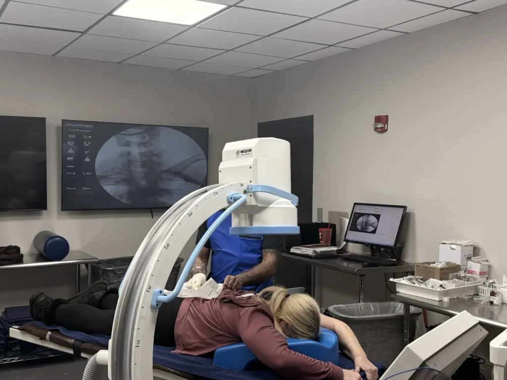
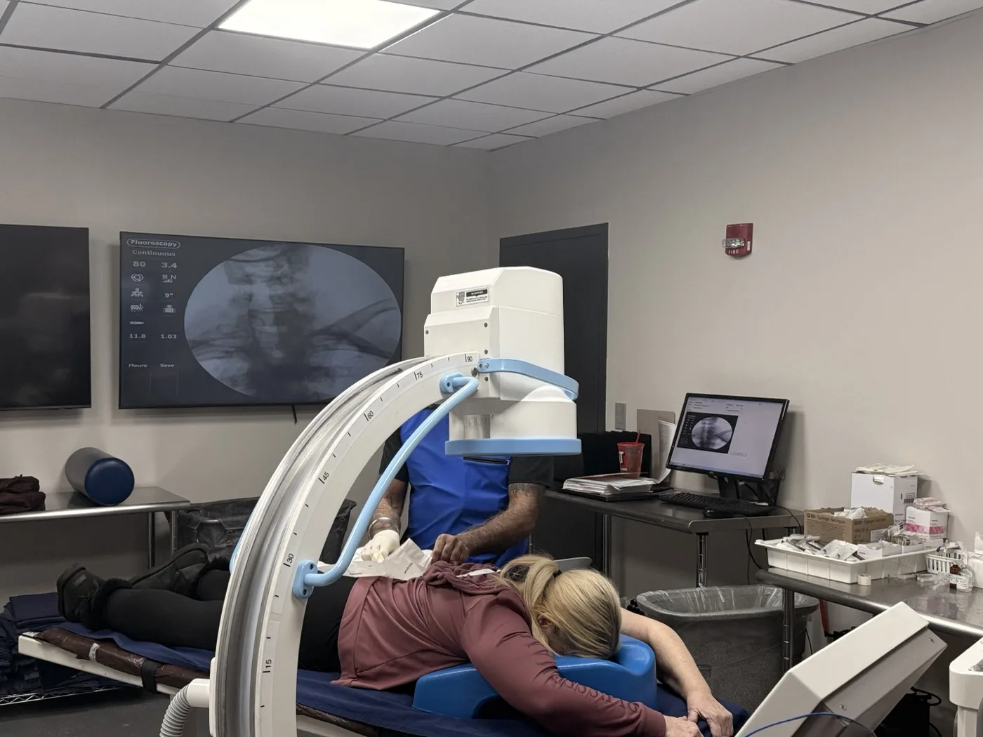
Image-guided injections are the gold standard for finding sacroiliac joint problems. The test records two things. First, the joint is filled and your familiar pain shows up. Second, the pain eases after infiltration of local anesthetic (numbing medicine).1 Both halves matter. Relief alone, without bringing on the matching pain, tells you less.
From most to least effective, the imaging choices are CT-guided, fluoroscopy-guided, and ultrasound-guided.1 The needle is aimed at the lower part of the joint. The joint is filled with only 1 to 2 mL of medicine. This is a small, tight space, not a cavity that can hold much.1
What a positive result actually buys you
A positive diagnostic response does not mean the joint is the only thing wrong. It means one pain source has been found and confirmed. So the next treatment can be aimed, not guessed. In a patient who has already cycled through treatments aimed at the low back, that is the whole point of the test.
Where a diagnostic block is used to confirm a target before a longer-lasting treatment, the logic is the same one used for facet joint pain and medial branch blocks. The procedure itself is described on the sacroiliac joint injection page. You can also see how it fits with the full range of pain treatments.
Risks worth asking about
Injections carry a risk of infection in the joint and of bleeding. Strict sterile technique and fine needles lower those risks.1 During image guidance, aiming at the lower part of the joint is essential. Radiofrequency denervation may cause numbness. Badly placed electrodes can damage sacral nerves and cause loss of bladder or bowel control, more pain, or limb weakness.1
These are not reasons to skip an evaluation. They are why image guidance and technique are not optional details. They are fair questions to ask before you agree to anything.
Frequently asked questions
How do doctors confirm the sacroiliac joint is the problem when my MRI is normal?
By numbing the joint under image guidance. The doctor checks whether filling the joint brings on your familiar pain and whether it is relieved by local anesthetic. That is a test with a result, not a guess from a picture. Why scans fall short here is covered in what imaging can and cannot show about pain.
My pain goes down my leg. Does that mean it is sciatica?
Not always. Sacroiliac joint pain has been reported to spread down the back or side of the thigh in 50% of cases, below the knee in 28%, and into the foot in 14%. So leg pain alone does not point to a nerve root. The other causes are compared in sciatica: disc, piriformis and cluneal nerve causes.
Is one positive provocative test enough to make the diagnosis?
No. Single tests hint at the answer but do not prove it. Accuracy improves when three or more provocation tests are positive. That exam is part of a first interventional visit, described on the pain management doctors in St. Louis page.
What are the risks of a sacroiliac joint injection?
Mainly infection and bleeding, which strict sterile technique and fine needles reduce. If radiofrequency denervation is considered later, numbness is possible. Badly placed electrodes can injure sacral nerves. Ask how image guidance is used, and see the sacroiliac joint injection page.
Do you see patients from Illinois for this?
Yes. The practice serves the St. Louis region across Missouri and Illinois. Both offices are listed on the locations page.
To discuss whether the sacroiliac joint is contributing to your own pain, request an appointment, call (314) 481-5000, or text (314) 886-5902.
Sources
- Raj MA, Ampat G, Varacallo MA. Sacroiliac Joint Pain. StatPearls. StatPearls Publishing; last updated August 14, 2023. NCBI Bookshelf NBK470299
Dr. Gurpreet Singh Padda, MD, MBA, MHP