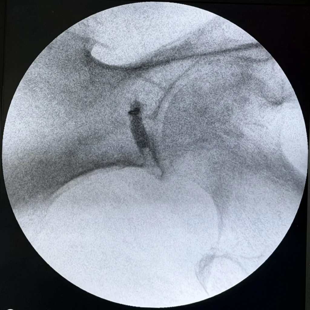
Fluoroscopy shows where a needle sits inside a piriformis muscle four to eight centimeters deep. The muscle lies right against the sciatic nerve, the nerve behind sciatica. No one can judge that depth by feel. The frame that proves placement is the contrast dye. It stretches along the body of the muscle and shows where the drug will go.
Most leg pain called sciatica gets blamed on a bulging disc. Much of the time, that is wrong. Say the spine has been scanned and treated, and the leg still hurts. Then the piriformis is one of the parts that has to be tested, not assumed. And testing it means putting medicine inside a muscle nobody can feel from the outside.
Piriformis syndrome is thought to cause somewhere between 0.3% and 6% of all low back and upper back-of-thigh pain. The United States sees roughly 40 million new sciatica-type back pain cases a year. So that still works out to about 2.4 million new cases a year.1 It is a small slice of a very large problem. The trouble is depth. The muscle sits four to eight centimeters deep in the buttock, right against the sciatic nerve. Once a needle goes under the skin, there is no reliable way to know where it is.
So we look. Below is a real X-ray sequence from a left piriformis injection done at the Woodson Road office. The frames are in the order they were taken. Nothing here is a diagram or a stock picture.
Why does a piriformis injection need X-ray or ultrasound guidance?
Two facts make landmark-only injection of the piriformis a poor idea. The first is depth. You can feel a trigger point in the trapezius (the upper back muscle). The piriformis is different. You cannot feel it with any precision through the big buttock muscle (the gluteus maximus) on top of it. The second is what it lies against. The sciatic nerve leaves the pelvis through a notch in the bone, right next to the muscle. In a fair share of people, it runs through the muscle instead of under it.
The StatPearls review of piriformis injection states the risk plainly. Accidental nerve irritation or injury is still a real risk of the procedure, particularly when needle localization is inaccurate. Fluoroscopic or ultrasound guidance improves needle placement and may reduce the risk of injury to nearby structures.1 That is the whole case for imaging. It is not a sales pitch.
Both fluoroscopy and ultrasound are used here for this injection. Where fluoroscopy is available, we prefer it. The images below make the reason obvious: contrast shows it better. Ultrasound shows you a needle. Contrast under fluoroscopy shows you where the drug is going to go.
The sequence, frame by frame
1. The baseline view
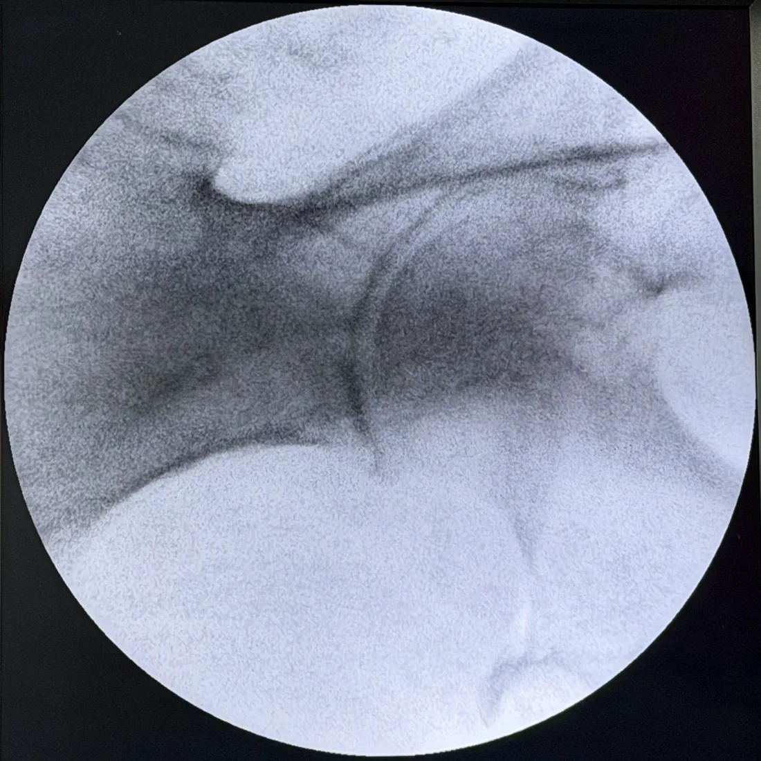
This frame does no medical work by itself. Its job is to set the view. Every later image will be read against it. It also confirms the bones sit where the anatomy says they should, before any needle goes in.
2. The needle advancing
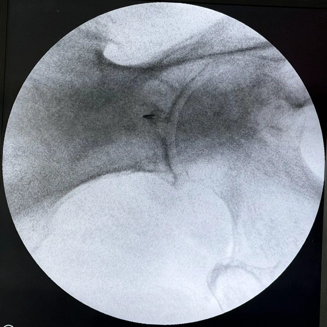
3. The tip at target
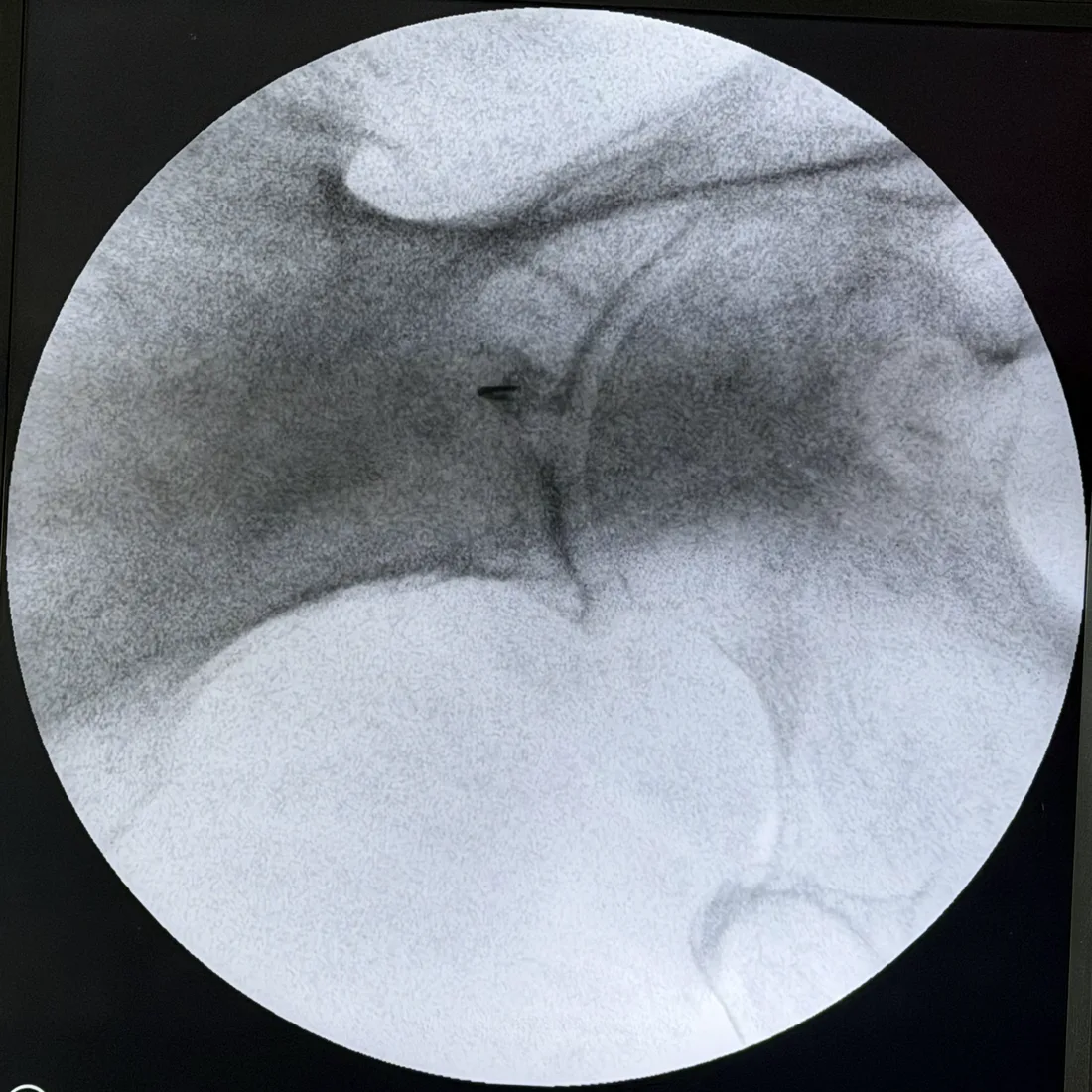
Here is where a landmark-only method would stop, because there is nothing more to check. The needle is where the doctor believes the muscle is. Whether that belief is right is still unknown.
4. Contrast — the frame that actually proves something
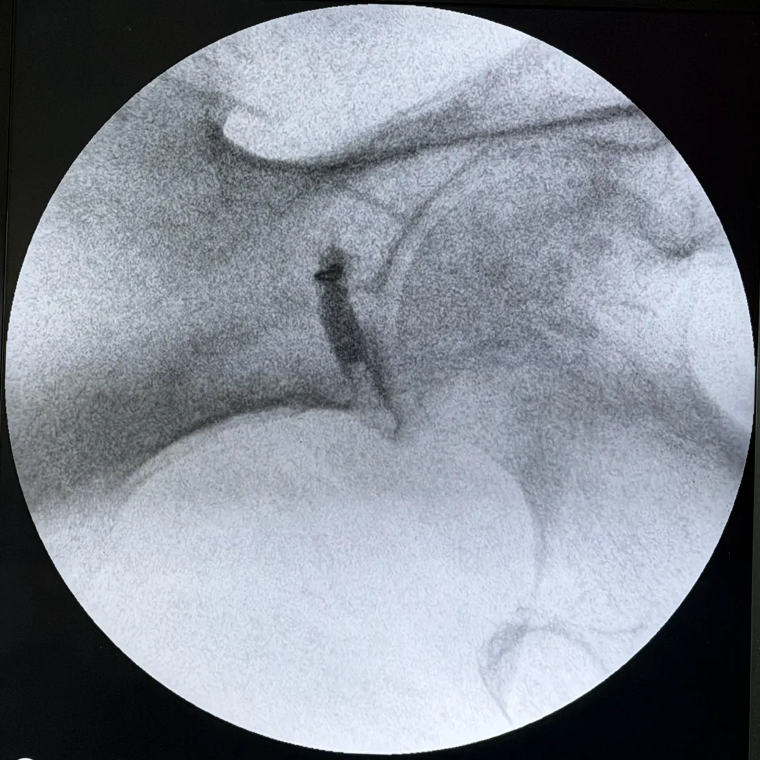
Should contrast be paired with fluoroscopy alone, with ultrasound, or with both? That is still an open question. The largest published look at it was a retrospective cohort of 78 patients and 109 procedures. Pain scores dropped the most in the group done with fluoroscopy and ultrasound together. Median procedure times were eight to eleven minutes across all three methods.3 The authors were careful. They called for prospective trials before treating that as settled.
This is the gap between a needle that looks correctly placed and an injection that is correctly placed. Contrast follows the path the drug will follow. Say it had slid into a layer between muscles, run toward the sciatic notch, or not spread at all. Then the needle would have been moved and a second contrast injection taken. All of that happens before a milligram of steroid or anesthetic goes anywhere.
5. After the injection
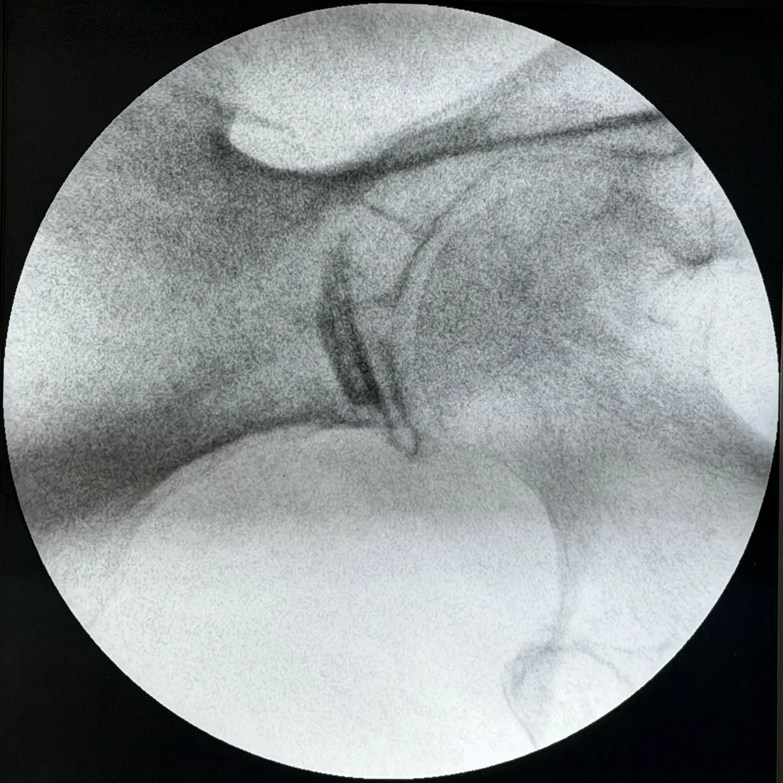
What contrast adds that needle position does not
Be precise here, because “image-guided” gets used loosely. There are three separate questions during a piriformis injection. They are not the same question:
- Where is the needle? Fluoroscopy answers this, live, in two dimensions.
- Where will the fluid go? Only contrast answers this. Needle position is a stand-in, and a poor one, because the tip can sit millimeters from the muscle and look the same on screen.
- Is this muscle the pain generator? Neither one answers this. Only how the patient responds over the next hours and days does.
Can imaging diagnose piriformis syndrome?
A clean contrast pattern is a technical win, not a diagnosis. The newest review of piriformis syndrome is blunt about the real problem. No well-accepted test confirms piriformis syndrome. No one knows how well the reported exam signs pick it up or rule it out. And in most published cases, the diagnosis seems to describe a muscle problem (a myofascial condition), not damage at one spot on a nerve (a focal neuropathy).2
The same review knocks down a myth worth retiring. People often say piriformis syndrome happens in those whose sciatic nerve runs through the muscle. The nerve does pierce the piriformis in roughly 16% of healthy people. But that rate is no different in people who have the syndrome. So any link to that anatomy is unclear.2 A body variant that is just as common in people with and without the problem does not explain the problem.
That is why the injection is used as a test as much as a treatment. It answers a question the imaging cannot.
How much radiation does a fluoroscopic piriformis injection use?
You can see the dose numbers in the frames above because the X-ray machine shows them and we did not crop them out. Across this sequence, the running total moved from 26.4 mGy to 28.8 mGy. We use fluoroscopy in short pulses to check position between moves. It is not left on the whole time. The total dose for a case like this is modest. It is a real dose, and it should be counted, not waved away. It is also why the procedure is done once, and done right. It is not repeated because nobody could confirm where the first one went.
Frequently asked questions
Does a piriformis injection hurt?
First we numb the skin and the path down to the muscle with local anesthetic. So the part most people dread is brief. Patients tend to notice something else more. They feel a deep pressure or a cramping ache as the fluid enters the muscle. It fades within a minute or two. We use no sedation. So you are awake and can tell us what you feel while we place the needle. That feedback helps us. It is not a side detail. Learn more: what actually happens during a piriformis injection.
Why is contrast used for a piriformis injection?
Contrast is the only thing in the room that shows where the fluid will actually go. A needle tip in the right spot on an X-ray still tells you nothing about spread. The tip could sit in fat, in a layer between muscles, or just outside the muscle. The image would look the same. We inject contrast first. It either takes the shape of the muscle or it does not. That answer comes before any drug is given. Learn more: the contrast frame in this image sequence.
Is fluoroscopy or ultrasound better for a piriformis injection?
We use both. We prefer fluoroscopy when it is available, because the contrast shows it better. Under fluoroscopy, we inject a small amount of contrast before any drug. It takes the shape of the muscle. So you see where the drug will actually spread, not just where the needle tip seems to sit. Ultrasound shows the needle and soft tissue live and adds no radiation. That makes it the right tool in some cases. But it does not create that record of spread. Published head-to-head data is thin. A 2022 retrospective cohort in Pain Physician looked at 78 patients and 109 procedures. Pain scores dropped the most in the group done with fluoroscopy and ultrasound together. The authors called for prospective trials before treating that as settled. The evidence agrees on one thing: image guidance of some kind beats none. Learn more: how image-guided procedures are performed here.
If the images look perfect, does that mean the injection will work?
No, and this is the most important limit to understand. A textbook contrast pattern proves the drug reached the piriformis muscle. It does not prove the piriformis muscle is what is causing your pain. No imaging test and no blood test confirms piriformis syndrome. The diagnosis is made by exam and history. The injection is part of how it gets tested. It is not proof the diagnosis was right. Learn more: what a failed piriformis injection tells you.
How long does a piriformis injection take?
The needle work itself is short. The largest published comparison looked at 78 patients and 109 procedures. Median procedure times ran eight to eleven minutes whether the injection was guided by fluoroscopy, ultrasound or both. That time includes the checking. The needle moves toward the muscle in small steps. Then contrast is injected to confirm the spread before any drug goes in.
Can a piriformis injection be done without ultrasound?
Yes, under fluoroscopy, which we prefer where it is available. What should not happen is an injection by feel alone. The muscle sits four to eight centimeters deep, right against the sciatic nerve. It cannot be felt through the buttock muscle on top of it. Fluoroscopic or ultrasound guidance improves needle placement and may reduce the risk of injury to nearby structures.
Does the sciatic nerve running through the piriformis cause piriformis syndrome?
Not by itself. The nerve passes through the piriformis muscle in roughly 16% of healthy people. That rate is no different in people who have piriformis syndrome. A variant that is just as common with and without the problem does not explain the problem. That is part of why no imaging or blood test confirms the diagnosis. It is also why the injection doubles as a test.
To discuss your own case, request an appointment through the appointment request form, call (314) 481-5000, or text (314) 886-5902. You can review the full range of pain treatments, read about the pain management doctors in St. Louis, or find both offices on the locations page.
Sources
- Chang A, Ly N, Varacallo MA. “Piriformis Injection.” StatPearls. Treasure Island (FL): StatPearls Publishing; 2023. NCBI Bookshelf NBK448193
- Lo JK, Robinson LR. “Piriformis syndrome.” Handbook of Clinical Neurology. 2024;201:203–226. doi:10.1016/B978-0-323-90108-6.00002-8
- Martiszus B, Hua S, Christiansen S, Ramsey K, Zusmer E. “A Novel Piriformis Injection Technique Utilizing Combined Fluoroscopy and Ultrasound — A Pilot Study.” Pain Physician. 2022;25(2):E365–E374. PMID 35322992
Dr. Gurpreet Singh Padda, MD, MBA, MHP